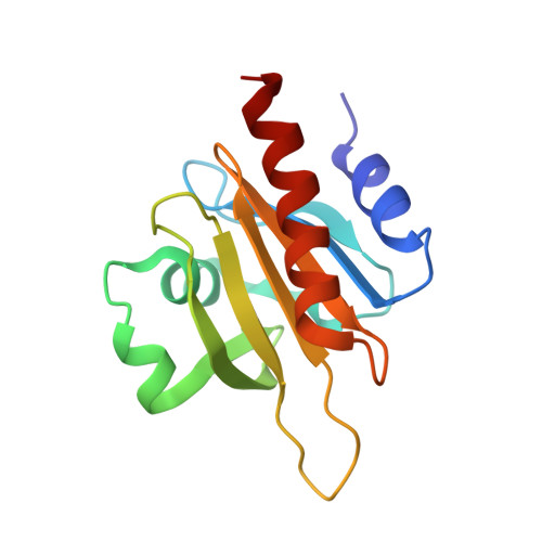
X-ray structure determination of human profilin II: A comparative structural analysis of human profilins.
Nodelman, I.M., Bowman, G.D., Lindberg, U., Schutt, C.E.(1999) J Mol Biology 294: 1271-1285
- PubMed: 10600384
- DOI: https://doi.org/10.1006/jmbi.1999.3318
- Primary Citation of Related Structures:
1D1J - PubMed Abstract:
Human profilins are multifunctional, single-domain proteins which directly link the actin microfilament system to a variety of signalling pathways via two spatially distinct binding sites. Profilin binds to monomeric actin in a 1:1 complex, catalyzes the exchange of the actin-bound nucleotide and regulates actin filament barbed end assembly. Like SH3 domains, profilin has a surface-exposed aromatic patch which binds to proline-rich peptides. Various multidomain proteins including members of the Ena/VASP and formin families localize profilin:actin complexes through profilin:poly-L-proline interactions to particular cytoskeletal locations (e.g. focal adhesions, cleavage furrows). Humans express a basic (I) and an acidic (II) isoform of profilin which exhibit different affinities for peptides and proteins rich in proline residues. Here, we report the crystallization and X-ray structure determination of human profilin II to 2.2 A. This structure reveals an aromatic extension of the previously defined poly-L-proline binding site for profilin I. In contrast to serine 29 of profilin I, tyrosine 29 in profilin II is capable of forming an additional stacking interaction and a hydrogen bond with poly-L-proline which may account for the increased affinity of the second isoform for proline-rich peptides. Differential isoform specificity for proline-rich proteins may be attributed to the differences in charged and hydrophobic residues in and proximal to the poly-L-proline binding site. The actin-binding face remains nearly identical with the exception of five amino acid differences. These observations are important for the understanding of the functional and structural differences between these two classes of profilin isoforms.
- Department of Molecular Biology, Henry H. Hoyt Laboratory, Princeton University, Princeton, NJ 08544, USA.
Organizational Affiliation: