1CQ2

NEUTRON STRUCTURE OF FULLY DEUTERATED SPERM WHALE MYOGLOBIN AT 2.0 ANGSTROM


Experimental Data Snapshot

  • Method: NEUTRON DIFFRACTION
  • Resolution: 2.00 Å
  • R-Value Free: 0.250 
  • R-Value Work: 0.160 
  • R-Value Observed: 0.160 

wwPDB Validation   3D Report Full Report


This is version 1.5 of the entry. See complete history


Literature

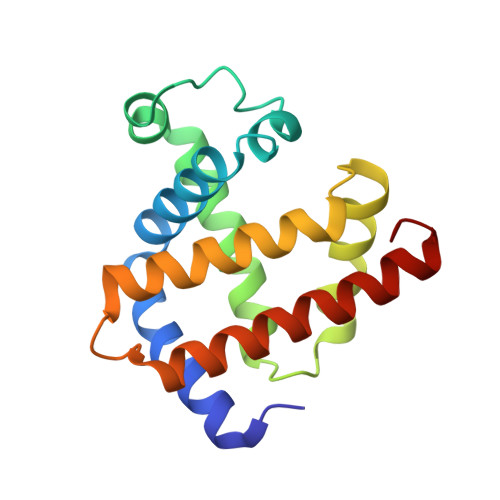
Enhanced visibility of hydrogen atoms by neutron crystallography on fully deuterated myoglobin.

Shu, F.Ramakrishnan, V.Schoenborn, B.P.

(2000) Proc Natl Acad Sci U S A 97: 3872-3877

  • DOI: https://doi.org/10.1073/pnas.060024697
  • Primary Citation of Related Structures:  
    1CQ2

  • PubMed Abstract: 

    Although hydrogens comprise half of the atoms in a protein molecule and are of great importance chemically and structurally, direct visualization of them by using crystallography is difficult. Neutron crystallography is capable of directly revealing the position of hydrogens, but its use on unlabeled samples faces certain technical difficulties: the large incoherent scattering of hydrogen results in background scattering that greatly reduces the signal to noise of the experiment. Moreover, whereas the scattering lengths of C, N, and O are positive, that of hydrogen is negative and about half the magnitude. This results in density for hydrogens being half as strong and close to the threshold of detection at 2.0-A resolution. Also, because of its opposite sign, there is a partial cancellation of the hydrogen density with that from neighboring atoms, which can lead to ambiguities in interpretation at medium resolution. These difficulties can be overcome by the use of deuterated protein, and we present here a neutron structure of fully deuterated myoglobin. The structure reveals a wealth of chemical information about the molecule, including the geometry of hydrogen bonding, states of protonation of histidines, and the location and geometry of water molecules at the surface of the protein. The structure also should be of broader interest because it will serve as a benchmark for molecular dynamics and energy minimization calculations and for comparison with NMR studies.


  • Organizational Affiliation

    Biology Department, Brookhaven National Laboratory, Upton, NY 11973, USA.


Macromolecules
Find similar proteins by:  (by identity cutoff)  |  3D Structure
Entity ID: 1
MoleculeChains Sequence LengthOrganismDetailsImage
MYOGLOBIN153Physeter catodonMutation(s): 0 
UniProt
Find proteins for P02185 (Physeter macrocephalus)
Explore P02185 
Go to UniProtKB:  P02185
Entity Groups  
Sequence Clusters30% Identity50% Identity70% Identity90% Identity95% Identity100% Identity
UniProt GroupP02185
Sequence Annotations
Expand
  • Reference Sequence
Small Molecules
Ligands 1 Unique
IDChains Name / Formula / InChI Key2D Diagram3D Interactions
HEM
Query on HEM

Download Ideal Coordinates CCD File 
B [auth A]PROTOPORPHYRIN IX CONTAINING FE
C34 H32 Fe N4 O4
KABFMIBPWCXCRK-RGGAHWMASA-L
Experimental Data & Validation

Experimental Data

  • Method: NEUTRON DIFFRACTION
  • Resolution: 2.00 Å
  • R-Value Free: 0.250 
  • R-Value Work: 0.160 
  • R-Value Observed: 0.160 
  • Space Group: P 1 21 1
Unit Cell:
Length ( Å )Angle ( ˚ )
a = 64.53α = 90
b = 30.91β = 105.82
c = 34.78γ = 90
Software Package:
Software NamePurpose
X-PLORmodel building
X-PLORrefinement
MADNESSdata reduction
CCP4data scaling
ROTAVATAdata scaling
X-PLORphasing

Structure Validation

View Full Validation Report


Entry History 

Deposition Data

Revision History  (Full details and data files)

  • Version 1.0: 1999-08-16
    Type: Initial release
  • Version 1.1: 2008-04-27
    Changes: Version format compliance
  • Version 1.2: 2011-07-13
    Changes: Non-polymer description, Version format compliance
  • Version 1.3: 2016-12-14
    Changes: Structure summary
  • Version 1.4: 2017-10-04
    Changes: Refinement description
  • Version 1.5: 2024-02-07
    Changes: Data collection, Database references, Derived calculations