Correlated disorder of the pure Pro22/Leu25 form of crambin at 150 K refined to 1.05-A resolution.
Yamano, A., Teeter, M.M.(1994) J Biol Chem 269: 13956-13965
- PubMed: 8188676
- Primary Citation of Related Structures:
1CNR - PubMed Abstract:
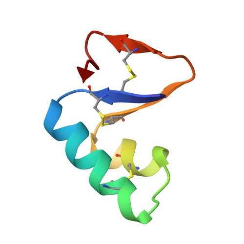
The high resolution crystal structure of crambin has been based on the crystals containing two sequence forms (the mixed form). Here, we report the crystal structure of the sequence isomer having Pro and Leu at residues 22 and 25 (the PL form). This elimination of the sequence heterogeneity resulted in a simpler structure which permits a more accurate modeling of protein disorder. In the observed disorder, the PL form structure and the mixed form structure have significant differences: 1) the disorder caused by the sequence heterogeneity (Pro2/Ser22, Leu/Ile25, Tyr29) is absent in the PL form; 2) Phe13 and Glu23 disordered in the mixed form have only one conformation in the PL form; and 3) Asn12 has multiple conformations in the PL form. During the study of disorder in the PL form structure, we found that conformational correlation can be inferred from a structure determined with Bragg's reflections by introducing fundamental stereochemical information, van der Waals contact (Gursky, O., Badger, J., Li, Y., and Caspar, D. L. D. (1992) Biophys. J. 63, 1210-1220), although an x-ray structure is an image averaged over a large number of copies and the period of data collection and does not carry direct evidence about correlations. The correlations among Thr2,Arg10, and Ile34 present the clearest example. The dimension of this correlation is comparable with the short-range (4-8 A) correlations in the atomic displacements concluded from the x-ray diffuse scattering experiments (Caspar, D. L. D., Clarage, J. B., Salunke, D. M., and Clarage, M. S. (1988) Nature 322, 659-662; Clarage, J. B., Clarage, M. S., Phillips, W. C., Sweet, R. M., and Caspar, D. L. D. (1992) Proteins 12, 145-157).
Organizational Affiliation:
Department of Chemistry, Merkert Chemistry Center, Boston College, Chestnut Hill, Massachusetts 02167.