Crystal structure of a hybrid between ribonuclease A and bovine seminal ribonuclease--the basic surface, at 2.0 A resolution.
Vatzaki, E.H., Allen, S.C., Leonidas, D.D., Trautwein-Fritz, K., Stackhouse, J., Benner, S.A., Acharya, K.R.(1999) Eur J Biochem 260: 176-182
- PubMed: 10091597
- DOI: https://doi.org/10.1046/j.1432-1327.1999.00142.x
- Primary Citation of Related Structures:
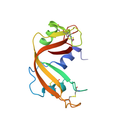
1B6V - PubMed Abstract:
A variant of bovine pancreatic ribonuclease A has been prepared with seven amino acid substitutions (Q55K, N62K, A64T, Y76K, S80R, E111G, N113K). These substitutions recreate in RNase A the basic surface found in bovine seminal RNase, a homologue of pancreatic RNase that diverged some 35 million years ago. Substitution of a portion of this basic surface (positions 55, 62, 64, 111 and 113) enhances the immunosuppressive activity of the RNase variant, activity found in native seminal RNase, while substitution of another portion (positions 76 and 80) attenuates the activity. Further, introduction of Gly at position 111 has been shown to increase the catalytic activity of RNase against double-stranded RNA. The variant and the wild-type (recombinant) protein were crystallized and their structures determined to a resolution of 2.0 A. Each of the mutated amino acids is seen in the electron density map. The main change observed in the mutant structure compared with the wild-type is the region encompassing residues 16-22, where the structure is more disordered. This loop is the region where the polypeptide chain of RNase A is cleaved by subtilisin to form RNase S, and undergoes conformational change to allow residues 1-20 of the RNase to swap between subunits in the covalent seminal RNase dimer.
- Department of Biology and Biochemistry, University of Bath, UK.
Organizational Affiliation: