Sequential 1H, 13C, and 15N NMR assignments and solution conformation of apokedarcidin.
Constantine, K.L., Colson, K.L., Wittekind, M., Friedrichs, M.S., Zein, N., Tuttle, J., Langley, D.R., Leet, J.E., Schroeder, D.R., Lam, K.S., Farmer III, B.T., Metzler, W.J., Bruccoleri, R.E., Mueller, L.(1994) Biochemistry 33: 11438-11452
- PubMed: 7918358
- DOI: https://doi.org/10.1021/bi00204a006
- Primary Citation of Related Structures:
1AKP - PubMed Abstract:
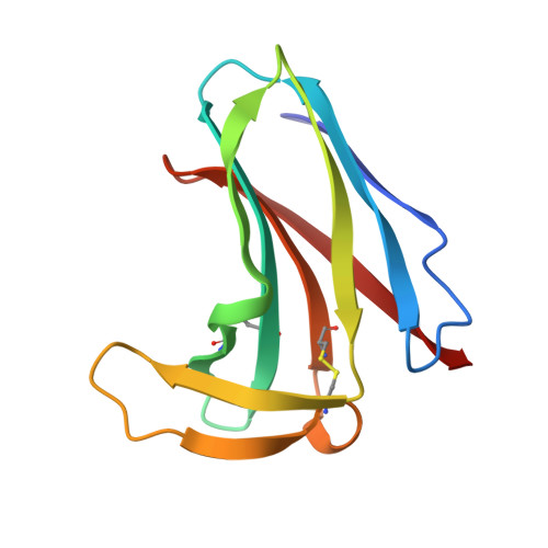
Kedarcidin is a recently discovered antitumor antibiotic chromoprotein. The solution conformation of the kedarcidin apoprotein (114 residues) has been characterized by heteronuclear multidimensional NMR spectroscopy. Sequence-specific backbone atom resonance assignments were obtained for a uniformly 13C/15N-enriched sample of apokedarcidin via a semiautomated analysis of 3D HNCACB, 3D CBCA-(CO)NH, 4D HNCAHA, 4D HN(CO)CAHA, 3D HBHA(CO)NH, and 3D HNHA(Gly) spectra. Side-chain assignments were subsequently obtained by analysis of (primarily) 3D HCCH-TOCSY and HCCH-COSY spectra. A qualitative analysis of the secondary structure is presented on the basis of 3J alpha NH coupling constants, deviations of 13C alpha and 13C beta chemical shifts from random coil values, and NOEs observed in 3D 15N- and 13C-edited NOESY-HSQC spectra. This analysis revealed a four-stranded antiparallel beta-sheet, a three-stranded antiparallel beta-sheet, and two two-standed antiparallel beta-sheets. The assignments of cross-peaks in the 3D NOESY spectra were assisted by reference to a preliminary model of apokedarcidin built using the program CONGEN starting from the X-ray structure of the homologous protein aponeocarzinostatin. An ensemble of 15 apokedarcidin solution structures has been generated by variable target function minimization (DIANA program) and refined by simulated annealing (X-PLOR program). The average backbone atom root-mean-square difference between the individual structures and the mean coordinates is 0.68 +/- 0.08 A. The overall fold of apokedarcidin is well-defined; it is composed of an immunoglobulin-like seven-stranded antiparallel beta-barrel and a subdomain containing two antiparallel beta-ribbons. Highly similar tertiary structures have been previously reported for the related proteins neocarzinostatin, macromomycin, and actinoxanthin. Important structural features are revealed, including the dimensions of the chromophore-binding pocket and the locations of side chains that are likely to be involved in chromophore stabilization.
Organizational Affiliation:
Bristol-Myers Squibb Pharmaceutical Research Institute, Princeton, New Jersey 08543-4000.