1W7E

NMR Ensemble OF Fasciclin-Like Protein From Rhodobacter sphaeroides


Experimental Data Snapshot

  • Method: SOLUTION NMR
  • Conformers Calculated: 100 
  • Conformers Submitted: 
  • Selection Criteria: 6 RANDOMLY CHOSEN FROM 30 LOWEST ENERGY 

wwPDB Validation   3D Report Full Report


This is version 1.2 of the entry. See complete history


Literature

Structure and Function of a Bacterial Fasciclin I Domain Protein Elucidates Function of Related Cell Adhesion Proteins Such as Tgfbip and Periostin.

Moody, R.G.Williamson, M.P.

(2013) FEBS Open Bio 3: 71

  • DOI: https://doi.org/10.1016/j.fob.2013.01.001
  • Primary Citation of Related Structures:  
    1W7D, 1W7E

  • PubMed Abstract: 

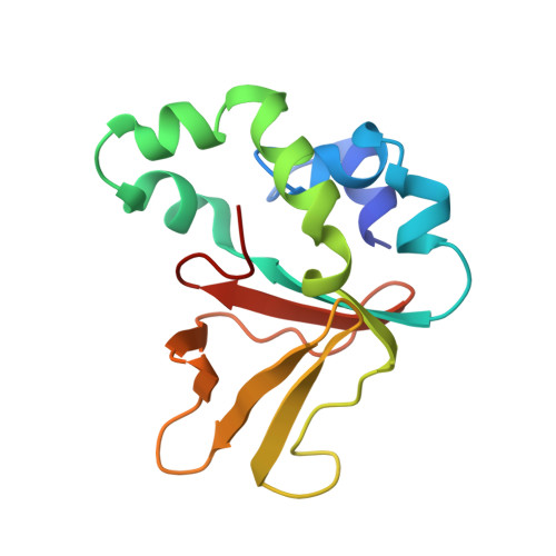
    Fasciclin I (FAS1) domains have important roles in cell adhesion, which are not understood despite many structural and functional studies. Examples of FAS1 domain proteins include TGFBIp (βig-h3) and periostin, which function in angiogenesis and development of cornea and bone, and are also highly expressed in cancer tissues. Here we report the structure of a single-domain bacterial fasciclin I protein, Fdp, in the free-living photosynthetic bacterium Rhodobacter sphaeroides, and show that it confers cell adhesion properties in vivo. A binding site is identified which includes the most highly conserved region and is adjacent to the N-terminus. By mapping this onto eukaryotic homologues, which all contain tandem FAS1 domains, it is concluded that the interaction site is normally buried in the dimer interface. This explains why corneal dystrophy mutations are concentrated in the C-terminal domain of TGFBIp and suggests new therapeutic approaches.


  • Organizational Affiliation

    Dept. of Molecular Biology and Biotechnology, Firth Court, Western Bank, University of Sheffield, Sheffield S10 2TN, UK.


Macromolecules
Find similar proteins by:  (by identity cutoff)  |  3D Structure
Entity ID: 1
MoleculeChains Sequence LengthOrganismDetailsImage
BETA-IG-H3/FASCICLIN137Cereibacter sphaeroidesMutation(s): 0 
UniProt
Find proteins for Q3IXZ6 (Cereibacter sphaeroides (strain ATCC 17023 / DSM 158 / JCM 6121 / CCUG 31486 / LMG 2827 / NBRC 12203 / NCIMB 8253 / ATH 2.4.1.))
Explore Q3IXZ6 
Go to UniProtKB:  Q3IXZ6
Entity Groups  
Sequence Clusters30% Identity50% Identity70% Identity90% Identity95% Identity100% Identity
UniProt GroupQ3IXZ6
Sequence Annotations
Expand
  • Reference Sequence
Experimental Data & Validation

Experimental Data

  • Method: SOLUTION NMR
  • Conformers Calculated: 100 
  • Conformers Submitted: 
  • Selection Criteria: 6 RANDOMLY CHOSEN FROM 30 LOWEST ENERGY 

Structure Validation

View Full Validation Report


Entry History 

Deposition Data

Revision History  (Full details and data files)

  • Version 1.0: 2006-03-08
    Type: Initial release
  • Version 1.1: 2013-06-19
    Changes: Atomic model, Database references, Derived calculations, Other, Refinement description, Source and taxonomy, Structure summary, Version format compliance
  • Version 1.2: 2014-07-30
    Changes: Database references