1R6Z

The Crystal Structure of the Argonaute2 PAZ domain (as a MBP fusion)


Experimental Data Snapshot

  • Method: X-RAY DIFFRACTION
  • Resolution: 2.80 Å
  • R-Value Free: 0.281 
  • R-Value Work: 0.227 

wwPDB Validation   3D Report Full Report


This is version 2.1 of the entry. See complete history


Literature

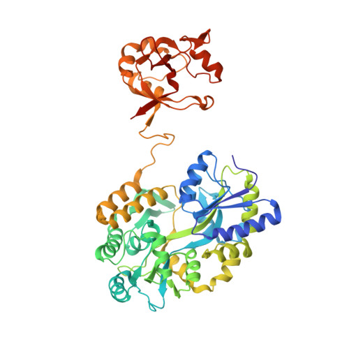
The crystal structure of the Argonaute2 PAZ domain reveals an RNA binding motif in RNAi effector complexes.

Song, J.J.Liu, J.Tolia, N.H.Schneiderman, J.Smith, S.K.Martienssen, R.A.Hannon, G.J.Joshua-Tor, L.

(2003) Nat Struct Biol 10: 1026-1032

  • DOI: https://doi.org/10.1038/nsb1016
  • Primary Citation of Related Structures:  
    1R6Z

  • PubMed Abstract: 

    RISC, the RNA-induced silencing complex, uses short interfering RNAs (siRNAs) or micro RNAs (miRNAs) to select its targets in a sequence-dependent manner. Key RISC components are Argonaute proteins, which contain two characteristic domains, PAZ and PIWI. PAZ is highly conserved and is found only in Argonaute proteins and Dicer. We have solved the crystal structure of the PAZ domain of Drosophila Argonaute2. The PAZ domain contains a variant of the OB fold, a module that often binds single-stranded nucleic acids. PAZ domains show low-affinity nucleic acid binding, probably interacting with the 3' ends of single-stranded regions of RNA. PAZ can bind the characteristic two-base 3' overhangs of siRNAs, indicating that although PAZ may not be a primary nucleic acid binding site in Dicer or RISC, it may contribute to the specific and productive incorporation of siRNAs and miRNAs into the RNAi pathway.


  • Organizational Affiliation

    Cold Spring Harbor Laboratory, Watson School of Biological Sciences, 1 Bungtown Road, Cold Spring Harbor, New York 11724, USA.


Macromolecules
Find similar proteins by:  (by identity cutoff)  |  3D Structure
Entity ID: 1
MoleculeChains Sequence LengthOrganismDetailsImage
Chimera of Maltose-binding periplasmic protein and Argonaute 2A [auth P],
B [auth A],
C [auth Z]
509Escherichia coliDrosophila melanogaster
This entity is chimeric
Mutation(s): 0 
Gene Names: MALEArgonaute 2 CG7439-PB
UniProt
Find proteins for P0AEX9 (Escherichia coli (strain K12))
Explore P0AEX9 
Go to UniProtKB:  P0AEX9
Find proteins for Q9VUQ5 (Drosophila melanogaster)
Explore Q9VUQ5 
Go to UniProtKB:  Q9VUQ5
Entity Groups  
Sequence Clusters30% Identity50% Identity70% Identity90% Identity95% Identity100% Identity
UniProt GroupsP0AEX9Q9VUQ5
Sequence Annotations
Expand
  • Reference Sequence
Oligosaccharides

Help

Entity ID: 2
MoleculeChains Length2D Diagram Glycosylation3D Interactions
alpha-D-glucopyranose-(1-4)-alpha-D-glucopyranoseD [auth B],
E [auth C],
F [auth D]
2N/A
Glycosylation Resources
GlyTouCan:  G07411ON
GlyCosmos:  G07411ON
Biologically Interesting Molecules (External Reference) 1 Unique
Experimental Data & Validation

Experimental Data

  • Method: X-RAY DIFFRACTION
  • Resolution: 2.80 Å
  • R-Value Free: 0.281 
  • R-Value Work: 0.227 
  • Space Group: P 65
Unit Cell:
Length ( Å )Angle ( ˚ )
a = 89.811α = 90
b = 89.811β = 90
c = 380.326γ = 120
Software Package:
Software NamePurpose
HKL-2000data collection
SCALEPACKdata scaling
AMoREphasing
CNSrefinement
HKL-2000data reduction

Structure Validation

View Full Validation Report


Entry History 

Deposition Data

Revision History  (Full details and data files)

  • Version 1.0: 2004-01-13
    Type: Initial release
  • Version 1.1: 2008-04-29
    Changes: Version format compliance
  • Version 1.2: 2011-07-13
    Changes: Derived calculations, Version format compliance
  • Version 1.3: 2017-08-16
    Changes: Refinement description, Source and taxonomy
  • Version 2.0: 2020-07-29
    Type: Remediation
    Reason: Carbohydrate remediation
    Changes: Atomic model, Data collection, Database references, Derived calculations, Non-polymer description, Structure summary
  • Version 2.1: 2023-08-23
    Changes: Data collection, Database references, Refinement description, Structure summary