1QFN

GLUTAREDOXIN-1-RIBONUCLEOTIDE REDUCTASE B1 MIXED DISULFIDE BOND


Experimental Data Snapshot

  • Method: SOLUTION NMR
  • Conformers Calculated: 50 
  • Conformers Submitted: 20 
  • Selection Criteria: LEAST TARGET FUNCTION 

wwPDB Validation   3D Report Full Report


This is version 1.4 of the entry. See complete history


Literature

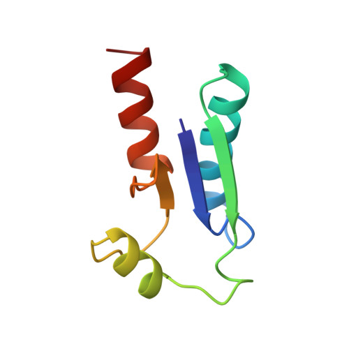
Binding specificity and mechanistic insight into glutaredoxin-catalyzed protein disulfide reduction.

Berardi, M.J.Bushweller, J.H.

(1999) J Mol Biol 292: 151-161

  • DOI: https://doi.org/10.1006/jmbi.1999.3067
  • Primary Citation of Related Structures:  
    1QFN

  • PubMed Abstract: 

    The reduction equivalents necessary for the ribonucleotide reductase (RNR)-catalyzed production of deoxyribonucleotides are provided by glutaredoxin (Grx) or thioredoxin (Trx). The initial location for transfer of reducing equivalents to RNR is located at the C terminus of the B1 subunit and involves the reduction of a disulfide between Cys754 and Cys759. We have used a 25-mer peptide corresponding to residues 737-761 of RNR B1 (C754-->S) to synthesize a stable mixed disulfide with Escherichia coli Grx-1 (C14-->S) resembling the structure of an intermediate in the reaction. The high-resolution solution structure of the mixed disulfide has been obtained by NMR with an RMSD of 0.56 A for all the backbone atoms of the protein and the well-defined portion of the peptide. The binding interactions responsible for specificity have been identified demonstrating the importance of electrostatic interactions in this system and providing a rationale for the specificity of the Grx-RNR interaction. The disulfide is buried in this complex, implying a solely intra-molecular mechanism of reduction in contrast to the previously determined structure of the glutathione complex where the disulfide was exposed; mutagenesis studies have shown the relevance of intermolecular reduction processes. Substantial conformational changes in the helices of the protein are associated with peptide binding which have significant mechanistic implications for protein disulfide reduction by glutaredoxins.


  • Organizational Affiliation

    Department of Molecular Physiology and Biological Physics, University of Virginia, Charlottesville, VA 22906-0011, USA.


Macromolecules
Find similar proteins by:  (by identity cutoff)  |  3D Structure
Entity ID: 1
MoleculeChains Sequence LengthOrganismDetailsImage
PROTEIN (GLUTAREDOXIN 1)85Escherichia coliMutation(s): 1 
UniProt
Find proteins for P68688 (Escherichia coli (strain K12))
Explore P68688 
Go to UniProtKB:  P68688
Entity Groups  
Sequence Clusters30% Identity50% Identity70% Identity90% Identity95% Identity100% Identity
UniProt GroupP68688
Sequence Annotations
Expand
  • Reference Sequence

Find similar proteins by:  Sequence   |   3D Structure  

Entity ID: 2
MoleculeChains Sequence LengthOrganismDetailsImage
PROTEIN (RIBONUCLEOSIDE-DIPHOSPHATE REDUCTASE 1)25Escherichia coliMutation(s): 0 
EC: 1.17.4.1
UniProt
Find proteins for P00452 (Escherichia coli (strain K12))
Explore P00452 
Go to UniProtKB:  P00452
Entity Groups  
Sequence Clusters30% Identity50% Identity70% Identity90% Identity95% Identity100% Identity
UniProt GroupP00452
Sequence Annotations
Expand
  • Reference Sequence
Experimental Data & Validation

Experimental Data

  • Method: SOLUTION NMR
  • Conformers Calculated: 50 
  • Conformers Submitted: 20 
  • Selection Criteria: LEAST TARGET FUNCTION 

Structure Validation

View Full Validation Report


Entry History 

Deposition Data

Revision History  (Full details and data files)

  • Version 1.0: 2000-01-01
    Type: Initial release
  • Version 1.1: 2008-04-26
    Changes: Version format compliance
  • Version 1.2: 2011-07-13
    Changes: Version format compliance
  • Version 1.3: 2021-11-03
    Changes: Database references, Derived calculations
  • Version 1.4: 2023-12-27
    Changes: Data collection