Solution structure of a trefoil-motif-containing cell growth factor, porcine spasmolytic protein.
Carr, M.D., Bauer, C.J., Gradwell, M.J., Feeney, J.(1994) Proc Natl Acad Sci U S A 91: 2206-2210
- PubMed: 8134374
- DOI: https://doi.org/10.1073/pnas.91.6.2206
- Primary Citation of Related Structures:
1PCP - PubMed Abstract:
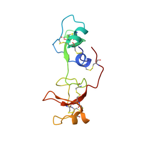
The porcine spasmolytic protein (pSP) is a 106-residue cell growth factor that typifies a family of eukaryotic proteins that contain at least one copy of an approximately 40-amino acid protein domain known as the trefoil motif. In fact, pSP contains two highly homologous trefoil domains. We have determined the complete three-dimensional solution structure of pSP by using a combination of two- and three-dimensional 1H NMR spectroscopy and distance geometry calculations. pSP is a relatively elongated molecule, consisting of two compact globular domains joined via a small interface. The protein's two trefoil domains adopt the same tertiary structure and contain a core C-terminal two-stranded antiparallel beta-sheet, preceded by a 6-residue helix that packs against the N-terminal beta-strand. The remainder of the protein backbone is taken up by two short loops that lie on either side of the beta-hairpin and are linked by an extended region that wraps around the C-terminal beta-strand. The topology of the protein backbone observed for the trefoil domains in pSP represents an unusual polypeptide fold. A striking feature of both trefoil domains is a surface patch formed from five conserved residues that have no obvious structural role. The two patches are located at the far ends of the protein molecule, and we propose that these residues form at least part of the receptor binding site, or sites, on pSP.
- Laboratory of Molecular Structure, National Institute for Medical Research, London, England.
Organizational Affiliation: