1NKN

VISUALIZING AN UNSTABLE COILED COIL: THE CRYSTAL STRUCTURE OF AN N-TERMINAL SEGMENT OF THE SCALLOP MYOSIN ROD


Experimental Data Snapshot

  • Method: X-RAY DIFFRACTION
  • Resolution: 2.50 Å
  • R-Value Free: 0.289 
  • R-Value Work: 0.243 
  • R-Value Observed: 0.247 

wwPDB Validation   3D Report Full Report


This is version 1.3 of the entry. See complete history


Literature

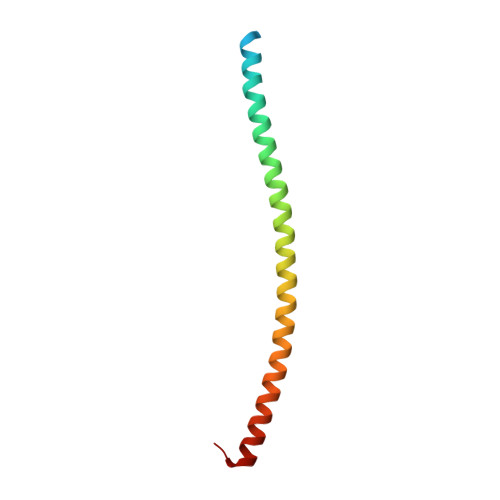
Visualization of an unstable coiled coil from the scallop myosin rod

Li, Y.Brown, J.H.Reshetnikova, L.Blazsek, A.Farkas, L.Nyitray, L.Cohen, C.

(2003) Nature 424: 341-345

  • DOI: https://doi.org/10.1038/nature01801
  • Primary Citation of Related Structures:  
    1NKN

  • PubMed Abstract: 

    Alpha-helical coiled coils in muscle exemplify simplicity and economy of protein design: small variations in sequence lead to remarkable diversity in cellular functions. Myosin II is the key protein in muscle contraction, and the molecule's two-chain alpha-helical coiled-coil rod region--towards the carboxy terminus of the heavy chain--has unusual structural and dynamic features. The amino-terminal subfragment-2 (S2) domains of the rods can swing out from the thick filament backbone at a hinge in the coiled coil, allowing the two myosin 'heads' and their motor domains to interact with actin and generate tension. Most of the S2 rod appears to be a flexible coiled coil, but studies suggest that the structure at the N-terminal region is unstable, and unwinding or bending of the alpha-helices near the head-rod junction seems necessary for many of myosin's functional properties. Here we show the physical basis of a particularly weak coiled-coil segment by determining the 2.5-A-resolution crystal structure of a leucine-zipper-stabilized fragment of the scallop striated-muscle myosin rod adjacent to the head-rod junction. The N-terminal 14 residues are poorly ordered; the rest of the S2 segment forms a flexible coiled coil with poorly packed core residues. The unusual absence of interhelical salt bridges here exposes apolar core atoms to solvent.


  • Organizational Affiliation

    Rosenstiel Basic Medical Sciences Research Center, Brandeis University, Waltham, Massachusetts 02454-9110, USA.


Macromolecules
Find similar proteins by:  (by identity cutoff)  |  3D Structure
Entity ID: 1
MoleculeChains Sequence LengthOrganismDetailsImage
S2N51-GCN4
A, B, C, D
89Argopecten irradiansSaccharomyces cerevisiae
This entity is chimeric
Mutation(s): 0 
UniProt
Find proteins for P03069 (Saccharomyces cerevisiae (strain ATCC 204508 / S288c))
Explore P03069 
Go to UniProtKB:  P03069
Find proteins for Q17042 (Argopecten irradians)
Explore Q17042 
Go to UniProtKB:  Q17042
Entity Groups  
Sequence Clusters30% Identity50% Identity70% Identity90% Identity95% Identity100% Identity
UniProt GroupsP03069Q17042
Sequence Annotations
Expand
  • Reference Sequence
Experimental Data & Validation

Experimental Data

  • Method: X-RAY DIFFRACTION
  • Resolution: 2.50 Å
  • R-Value Free: 0.289 
  • R-Value Work: 0.243 
  • R-Value Observed: 0.247 
  • Space Group: P 21 21 21
Unit Cell:
Length ( Å )Angle ( ˚ )
a = 54.034α = 90
b = 73.303β = 90
c = 102.975γ = 90
Software Package:
Software NamePurpose
DENZOdata reduction
SCALEPACKdata scaling
AMoREphasing
CNSrefinement

Structure Validation

View Full Validation Report


Entry History 

Deposition Data

Revision History  (Full details and data files)

  • Version 1.0: 2003-07-22
    Type: Initial release
  • Version 1.1: 2008-04-29
    Changes: Version format compliance
  • Version 1.2: 2011-07-13
    Changes: Version format compliance
  • Version 1.3: 2024-02-14
    Changes: Data collection, Database references