1NAN

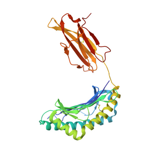
MCH CLASS I H-2KB MOLECULE COMPLEXED WITH PBM1 PEPTIDE


Experimental Data Snapshot

  • Method: X-RAY DIFFRACTION
  • Resolution: 2.30 Å
  • R-Value Free: 0.279 
  • R-Value Work: 0.224 
  • R-Value Observed: 0.229 

Starting Model: experimental
View more details

wwPDB Validation   3D Report Full Report


This is version 1.3 of the entry. See complete history


Literature

CDR3 loop flexibility contributes to the degeneracy of TCR recognition

Reiser, J.-B.Darnault, C.Gregoire, C.Mosser, T.Mazza, G.Kearnay, A.van der Merwe, P.A.Fontecilla-Camps, J.C.Housset, D.Malissen, B.

(2003) Nat Immunol 4: 241-247

  • DOI: https://doi.org/10.1038/ni891
  • Primary Citation of Related Structures:  
    1NAM, 1NAN

  • PubMed Abstract: 

    T cell receptor (TCR) binding degeneracy lies at the heart of several physiological and pathological phenomena, yet its structural basis is poorly understood. We determined the crystal structure of a complex involving the BM3.3 TCR and an octapeptide (VSV8) bound to the H-2K(b) major histocompatibility complex molecule at a 2.7 A resolution, and compared it with the BM3.3 TCR bound to the H-2K(b) molecule loaded with a peptide that has no primary sequence identity with VSV8. Comparison of these structures showed that the BM3.3 TCR complementarity-determining region (CDR) 3alpha could undergo rearrangements to adapt to structurally different peptide residues. Therefore, CDR3 loop flexibility helps explain TCR binding cross-reactivity.


  • Organizational Affiliation

    Laboratoire de Cristallographie et Cristallogenèse des Protéines, Institut de Biologie Structurale J.-P. Ebel, CEA-CNRS-UJF, 41 rue Jules Horowitz, F-38027 Grenoble Cedex 1, France.


Macromolecules
Find similar proteins by:  (by identity cutoff)  |  3D Structure
Entity ID: 1
MoleculeChains Sequence LengthOrganismDetailsImage
H-2 class I histocompatibility antigen, K-B alpha chainA [auth H],
B [auth L]
278Mus musculusMutation(s): 0 
Gene Names: H2-K
UniProt
Find proteins for P01901 (Mus musculus)
Explore P01901 
Go to UniProtKB:  P01901
Entity Groups  
Sequence Clusters30% Identity50% Identity70% Identity90% Identity95% Identity100% Identity
UniProt GroupP01901
Sequence Annotations
Expand
  • Reference Sequence
Find similar proteins by:  (by identity cutoff)  |  3D Structure
Entity ID: 2
MoleculeChains Sequence LengthOrganismDetailsImage
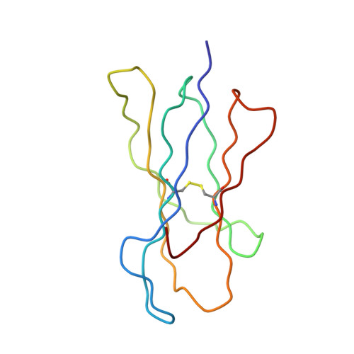
Beta-2-microglobulinC [auth P],
D [auth I]
99Mus musculusMutation(s): 0 
Gene Names: B2M
UniProt & NIH Common Fund Data Resources
Find proteins for P01887 (Mus musculus)
Explore P01887 
Go to UniProtKB:  P01887
IMPC:  MGI:88127
Entity Groups  
Sequence Clusters30% Identity50% Identity70% Identity90% Identity95% Identity100% Identity
UniProt GroupP01887
Sequence Annotations
Expand
  • Reference Sequence

Find similar proteins by:  Sequence   |   3D Structure  

Entity ID: 3
MoleculeChains Sequence LengthOrganismDetailsImage
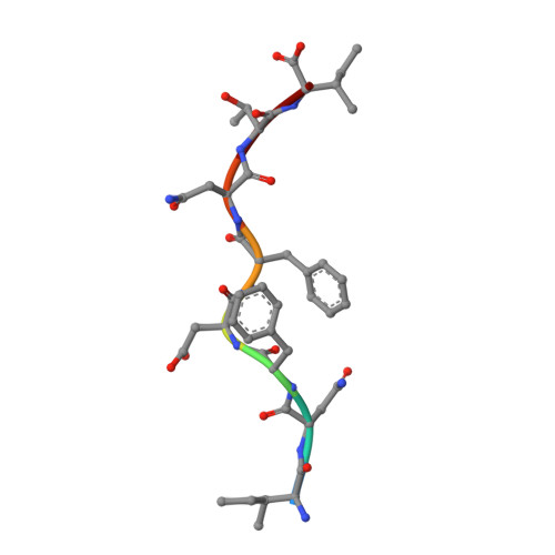
pBM1 peptideE [auth M],
F [auth Q]
8N/AMutation(s): 0 
UniProt
Find proteins for Q8CDD8 (Mus musculus)
Explore Q8CDD8 
Go to UniProtKB:  Q8CDD8
Entity Groups  
UniProt GroupQ8CDD8
Sequence Annotations
Expand
  • Reference Sequence
Experimental Data & Validation

Experimental Data

  • Method: X-RAY DIFFRACTION
  • Resolution: 2.30 Å
  • R-Value Free: 0.279 
  • R-Value Work: 0.224 
  • R-Value Observed: 0.229 
  • Space Group: P 1 21 1
Unit Cell:
Length ( Å )Angle ( ˚ )
a = 65.981α = 90
b = 88.614β = 111.6
c = 89.174γ = 90
Software Package:
Software NamePurpose
MOSFLMdata reduction
SCALAdata scaling
AMoREphasing
REFMACrefinement
CCP4data scaling

Structure Validation

View Full Validation Report


Entry History 

Revision History  (Full details and data files)

  • Version 1.0: 2003-03-11
    Type: Initial release
  • Version 1.1: 2008-04-28
    Changes: Version format compliance
  • Version 1.2: 2011-07-13
    Changes: Version format compliance
  • Version 1.3: 2023-08-16
    Changes: Data collection, Database references, Refinement description