1FZ3

METHANE MONOOXYGENASE HYDROXYLASE, FORM III SOAK AT PH 6.2 (0.1 M PIPES)


Experimental Data Snapshot

  • Method: X-RAY DIFFRACTION
  • Resolution: 2.03 Å
  • R-Value Free: 0.228 
  • R-Value Work: 0.196 
  • R-Value Observed: 0.196 

wwPDB Validation   3D Report Full Report


This is version 1.3 of the entry. See complete history


Literature

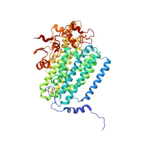
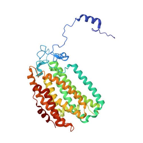
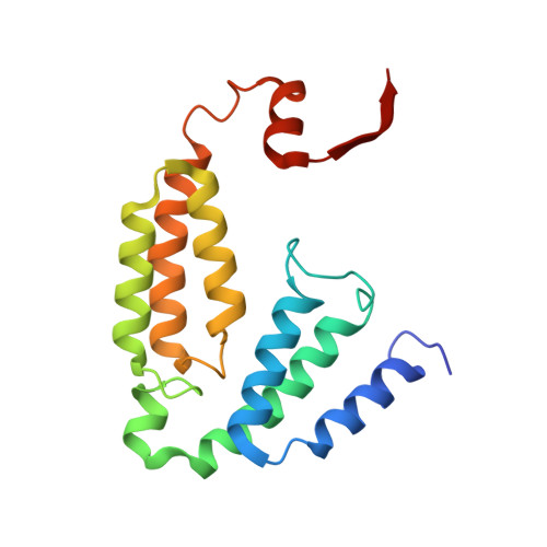
Crystal structures of the soluble methane monooxygenase hydroxylase from Methylococcus capsulatus (Bath) demonstrating geometrical variability at the dinuclear iron active site.

Whittington, D.A.Lippard, S.J.

(2001) J Am Chem Soc 123: 827-838

  • DOI: https://doi.org/10.1021/ja003240n
  • Primary Citation of Related Structures:  
    1FYZ, 1FZ0, 1FZ1, 1FZ2, 1FZ3, 1FZ4, 1FZ5

  • PubMed Abstract: 

    The oxidation of methane to methanol is performed at carboxylate-bridged dinuclear iron centers in the soluble methane monooxygenase hydroxylase (MMOH). Previous structural studies of MMOH, and the related R2 subunit of ribonucleotide reductase, have demonstrated the occurrence of carboxylate shifts involving glutamate residues that ligate the catalytic iron atoms. These shifts are thought to have important mechanistic implications. Recent kinetic and theoretical studies have also emphasized the importance of hydrogen bonding and pH effects at the active site. We report here crystal structures of MMOH from Methylococcus capsulatus (Bath) in the diiron(II), diiron(III), and mixed-valent Fe(II)Fe(III) oxidation states, and at pH values of 6.2, 7.0, and 8.5. These structures were investigated in an effort to delineate the range of possible motions at the MMOH active site and to identify hydrogen-bonding interactions that may be important in understanding catalysis by the enzyme. Our results present the first view of the diiron center in the mixed-valent state, and they indicate an increased lability for ferrous ions in the enzyme. Alternate conformations of Asn214 near the active site according to redox state and a distortion in one of the alpha-helices adjacent to the metal center in the diiron(II) state have also been identified. These changes alter the surface of the protein in the vicinity of the catalytic core and may have implications for small-molecule accessibility to the active site and for protein component interactions in the methane monooxygenase system. Collectively, these results help to explain previous spectroscopic observations and provide new insight into catalysis by the enzyme.


  • Organizational Affiliation

    Department of Chemistry, Massachusetts Institute of Technology, Cambridge, Massachusetts 02139, USA.


Macromolecules
Find similar proteins by:  (by identity cutoff)  |  3D Structure
Entity ID: 1
MoleculeChains Sequence LengthOrganismDetailsImage
METHANE MONOOXYGENASE COMPONENT A, ALPHA CHAIN
A, B
527Methylococcus capsulatusMutation(s): 0 
EC: 1.14.13.25
UniProt
Find proteins for P22869 (Methylococcus capsulatus (strain ATCC 33009 / NCIMB 11132 / Bath))
Explore P22869 
Go to UniProtKB:  P22869
Entity Groups  
Sequence Clusters30% Identity50% Identity70% Identity90% Identity95% Identity100% Identity
UniProt GroupP22869
Sequence Annotations
Expand
  • Reference Sequence
Find similar proteins by:  (by identity cutoff)  |  3D Structure
Entity ID: 2
MoleculeChains Sequence LengthOrganismDetailsImage
METHANE MONOOXYGENASE COMPONENT A, BETA CHAIN
C, D
389Methylococcus capsulatusMutation(s): 0 
EC: 1.14.13.25
UniProt
Find proteins for P18798 (Methylococcus capsulatus (strain ATCC 33009 / NCIMB 11132 / Bath))
Explore P18798 
Go to UniProtKB:  P18798
Entity Groups  
Sequence Clusters30% Identity50% Identity70% Identity90% Identity95% Identity100% Identity
UniProt GroupP18798
Sequence Annotations
Expand
  • Reference Sequence
Find similar proteins by:  (by identity cutoff)  |  3D Structure
Entity ID: 3
MoleculeChains Sequence LengthOrganismDetailsImage
METHANE MONOOXYGENASE COMPONENT A, GAMMA CHAIN
E, F
170Methylococcus capsulatusMutation(s): 0 
EC: 1.14.13.25
UniProt
Find proteins for P11987 (Methylococcus capsulatus (strain ATCC 33009 / NCIMB 11132 / Bath))
Explore P11987 
Go to UniProtKB:  P11987
Entity Groups  
Sequence Clusters30% Identity50% Identity70% Identity90% Identity95% Identity100% Identity
UniProt GroupP11987
Sequence Annotations
Expand
  • Reference Sequence
Experimental Data & Validation

Experimental Data

  • Method: X-RAY DIFFRACTION
  • Resolution: 2.03 Å
  • R-Value Free: 0.228 
  • R-Value Work: 0.196 
  • R-Value Observed: 0.196 
  • Space Group: P 21 21 21
Unit Cell:
Length ( Å )Angle ( ˚ )
a = 71.37α = 90
b = 172.12β = 90
c = 221.39γ = 90
Software Package:
Software NamePurpose
CNSrefinement
DENZOdata reduction
SCALEPACKdata scaling
CNSphasing

Structure Validation

View Full Validation Report


Entry History 

Deposition Data

Revision History  (Full details and data files)

  • Version 1.0: 2001-02-21
    Type: Initial release
  • Version 1.1: 2008-04-27
    Changes: Version format compliance
  • Version 1.2: 2011-07-13
    Changes: Version format compliance
  • Version 1.3: 2024-02-07
    Changes: Data collection, Database references, Derived calculations