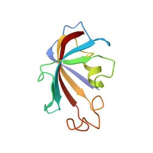
FK-506-binding protein: three-dimensional structure of the complex with the antagonist L-685,818.
Becker, J.W., Rotonda, J., McKeever, B.M., Chan, H.K., Marcy, A.I., Wiederrecht, G., Hermes, J.D., Springer, J.P.(1993) J Biological Chem 268: 11335-11339
- PubMed: 7684380
- Primary Citation of Related Structures:
1FKD, 2FKE - PubMed Abstract:
L-685,818 differs only slightly in structure from the immunosuppressive drug FK-506, and both compounds bind with comparable affinity to the 12-kDa FK-506-binding protein (FKBP12), the major intracellular receptor for the drug. Despite these similarities, L-685,818 is a potent antagonist of both the immunosuppressive and toxic effects of the drug. Here, we present a structural analysis of this problem. Although FK-506 and L-685,818 differ greatly in pharmacology, we have found that the three-dimensional structures of their complexes with FKBP12 are essentially identical. Approximately half of each ligand is in contact with the receptor protein, and half is exposed to solvent; the exposed region includes the two sites where the compounds differ. These results indicate that the profound differences in the pharmacology of these two compounds are not caused by any difference in their interaction with FKBP12. Rather, these effects arise because relatively minor changes in the exposed part of a bound ligand have a strong effect on how FKBP12-ligand complexes interact with calcineurin, their putative intracellular target. In addition, FK-506 complexes with FKBP12 proteins from several species all inhibit mammalian calcineurin. Analysis of the three-dimensional structure of the complex with respect to residues conserved among these proteins suggests a small number of surface residues near the bound ligands that may play a critical role in interactions between the protein-drug complex and calcineurin.
- Department of Biophysical Chemistry, Merck Research Laboratories, Rahway, New Jersey 07065-0900.
Organizational Affiliation: