Solution structure of the DNA-binding domain of a human papillomavirus E2 protein: evidence for flexible DNA-binding regions.
Liang, H., Petros, A.M., Meadows, R.P., Yoon, H.S., Egan, D.A., Walter, K., Holzman, T.F., Robins, T., Fesik, S.W.(1996) Biochemistry 35: 2095-2103
- PubMed: 8652551
- DOI: https://doi.org/10.1021/bi951932w
- Primary Citation of Related Structures:
1DHM - PubMed Abstract:
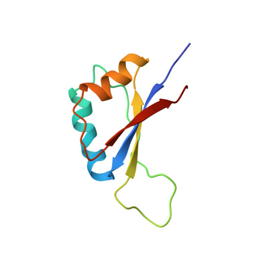
The three-dimensional structure of the DNA-binding domain of the E2 protein from human papillomavirus-31 was determined by using multidimensional heteronuclear nuclear magnetic resonance (NMR) spectroscopy. A total of 1429 NMR-derived distance and dihedral angle restraints were obtained for each of the 83-residue subunits of this symmetric dimer. The average root mean square deviations of 20 structures calculated using a distance geometry-simulated annealing protocol are 0.59 and 0.90 angstroms for the backbone and all heavy atoms, respectively, for residues 2-83. The structure of the human virus protein free in solution consists of an eight-stranded beta-barrel and two pairs of alpha-helices. Although the overall fold of the protein is similar to the crystal structure of the bovine papillomavirus-1 E2 protein when complexed to DNA, several small but interesting differences were observed between these two structures at the subunit interface. In addition, a beta-hairpin that contacts DNA in the crystal structure of the protein-DNA complex is disordered in the NMR structures, and steady-state 1H-15N heteronuclear NOE measurements indicate that this region is highly mobile in the absence of DNA. The recognition helix also appears to be flexible, as evidenced by fast amide exchange rates. This phenomenon has also been observed for a number of other DNA-binding proteins and may constitute a common theme in protein/DNA recognition.
- Pharmaceutical Discovery Division, Abbott Laboratories, Abbott Park, Illinois 60064, USA.
Organizational Affiliation: