On the role of the cis-proline residue in the active site of DsbA.
Charbonnier, J.B., Belin, P., Moutiez, M., Stura, E.A., Quemeneur, E.(1999) Protein Sci 8: 96-105
- PubMed: 10210188
- DOI: https://doi.org/10.1110/ps.8.1.96
- Primary Citation of Related Structures:
1BQ7 - PubMed Abstract:
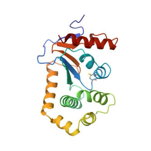
In addition to the Cys-Xaa-Xaa-Cys motif at position 30-33, DsbA, the essential catalyst for disulfide bond formation in the bacterial periplasm shares with other oxidoreductases of the thioredoxin family a cis-proline in proximity of the active site residues. In the variant DsbA(P151A), this residue has been changed to an alanine, an almost isosteric residue which is not disposed to adopt the cis conformation. The substitution strongly destabilized the structure of DsbA, as determined by the decrease in the free energy of folding. The pKa of the thiol of Cys30 was only marginally decreased. Although in vivo the variant appeared to be correctly oxidized, it exhibited an activity less than half that of the wild-type enzyme with respect to the folding of alkaline phosphatase, used as a reporter of the disulfide bond formation in the periplasm. DsbA(P151A) crystallized in a different crystal form from the wild-type protein, in space group P2(1) with six molecules in the asymmetric unit. Its X-ray structure was determined to 2.8 A resolution. The most significant conformational changes occurred at the active site. The loop 149-152 adopted a new backbone conformation with Ala151 in a trans conformation. This rearrangement resulted in the loss of van der Waals interactions between this loop and the disulfide bond. His32 from the Cys-Xaa-Xaa-Cys sequence presented in four out of six molecules in the asymmetric unit a gauche conformation not observed in the wild-type protein. The X-ray structure and folding studies on DsbA(P151A) were consistent with the cis-proline playing a major role in the stabilization of the protein. A role for the positioning of the substrate is discussed. These important properties for the enzyme function might explain the conservation of this residue in DsbA and related proteins possessing the thioredoxin fold.
Organizational Affiliation:
CEA, Département d'Ingénierie et d'Etudes des Protéines, Gif-sur-Yvette, France.