12AO | pdb_000012ao

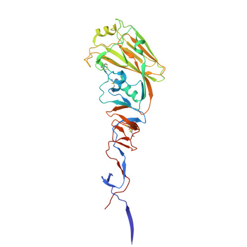
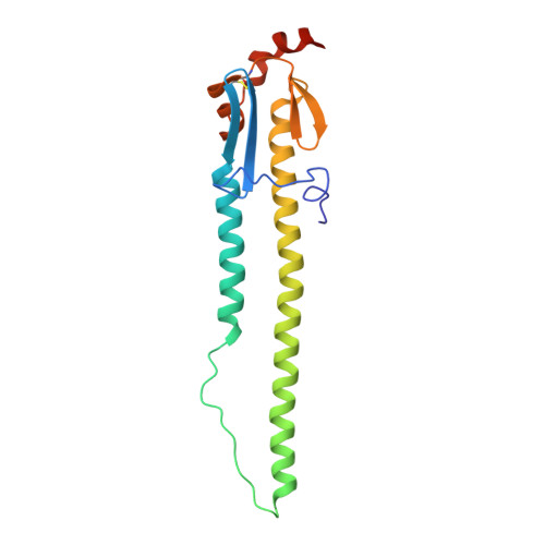
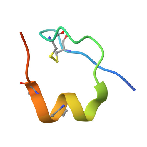
Crystal structure of the A/Puerto Rico/8/1934 (H1N1) influenza virus hemagglutinin in complex with fusion inhibitor peptide DP142014


Experimental Data Snapshot

  • Method: X-RAY DIFFRACTION
  • Resolution: 2.85 Å
  • R-Value Free: 
    0.247 (Depositor), 0.243 (DCC) 
  • R-Value Work: 
    0.208 (Depositor), 0.208 (DCC) 
  • R-Value Observed: 
    0.210 (Depositor) 

Starting Model: experimental
View more details

wwPDB Validation 3D Report Full Report

Validation slider image for 12AO

Ligand Structure Quality Assessment 


This is version 1.1 of the entry. See complete history. 

Literature

Antibody Hotspot-Centric de Novo Design of Peptide Inhibitors of Influenza A Virus Membrane Fusion.

Kadam, R.U., Juraszek, J., Branduardi, D., van Ameijde, J., Garg, D., Vermond, J., Jongeneelen, M., Tang, C., Vogels, R., van Dongen, M.J.P., Brandenburg, B., Friesen, R.H.E., Wilson, I.A.

(2026) ACS Med Chem Lett 17: 2191-2196

  • DOI: https://doi.org/10.1021/acsmedchemlett.6c00321
  • Primary Citation Related Structures: 
    12AO

  • PubMed Abstract: 

    Here we report the applicability of a hotspot-centric approach for de novo computational design of l-peptides based on crystal structures of antigen-antibody complexes. Broadly neutralizing antibodies (bnAbs) FI6v3 and CR9114, which target influenza A virus hemagglutinin (HA), guided the peptide design. Disembodied hotspot residues from these bnAbs were selected and anchored onto known peptide scaffolds (≤35 amino acids) from the Protein Data Bank (PDB). The top-scoring designs were subsequently synthesized, folded, tested in vitro , and structurally characterized in complex with HA. The designed peptides demonstrate structural and functional mimicry by recapitulating the binding mode of the bnAbs on HA and act as inhibitors of low-pH-dependent conformational transitions in HA that facilitate membrane fusion. These findings demonstrate that hotspot-guided design strategy, previously applied to proteins and d-peptides, can be effectively extended to l-peptide scaffolds, enabling the development of functional HA inhibitors informed by key residues at antigen-antibody interfaces.


  • Organizational Affiliation: 
    • Department of Integrative Structural and Computational Biology, The Scripps Research Institute, La Jolla, California 92037, United States.

Macromolecule Content 

  • Total Structure Weight: 61.88 kDa 
  • Atom Count: 4,217 
  • Modeled Residue Count: 517 
  • Deposited Residue Count: 532 
  • Unique protein chains: 3

Macromolecules

Find similar proteins by:|  3D Structure
Entity ID: 1
MoleculeChains  Sequence LengthOrganismDetailsImage
Hemagglutinin326Influenza A virus (A/Puerto Rico/8/1934(H1N1))Mutation(s): 0 
Gene Names: HA
UniProt
Find proteins for P03452 (Influenza A virus (strain A/Puerto Rico/8/1934 H1N1))
Explore P03452 
Go to UniProtKB:  P03452
Entity Groups
Sequence Clusters30% Identity50% Identity70% Identity90% Identity95% Identity100% Identity
UniProt GroupP03452
Glycosylation
Glycosylation Sites: 3
Sequence Annotations
Expand
Reference Sequence
Find similar proteins by:|  3D Structure
Entity ID: 2
MoleculeChains  Sequence LengthOrganismDetailsImage
Hemagglutinin HA2 chain176Influenza A virus (A/Puerto Rico/8/1934(H1N1))Mutation(s): 0 
Gene Names: HA
UniProt
Find proteins for P03452 (Influenza A virus (strain A/Puerto Rico/8/1934 H1N1))
Explore P03452 
Go to UniProtKB:  P03452
Entity Groups
Sequence Clusters30% Identity50% Identity70% Identity90% Identity95% Identity100% Identity
UniProt GroupP03452
Glycosylation
Glycosylation Sites: 1
Sequence Annotations
Expand
Reference Sequence
Find similar proteins by:|  3D Structure
Entity ID: 3
MoleculeChains  Sequence LengthOrganismDetailsImage
Synthetic peptide30PapillomaviridaeMutation(s): 0 
Entity Groups
Sequence Clusters30% Identity50% Identity70% Identity90% Identity95% Identity100% Identity
Sequence Annotations
Expand
Reference Sequence

Oligosaccharides

Help  
Entity ID: 4
MoleculeChains Length2D Diagram GlycosylationD Interactions
2-acetamido-2-deoxy-beta-D-glucopyranose-(1-4)-2-acetamido-2-deoxy-beta-D-glucopyranoseD [auth P],
E [auth D]
2N-Glycosylation
Glycosylation Resources
GlyTouCan: G42666HT
GlyCosmos: G42666HT
GlyGen: G42666HT

Small Molecules

Modified Residues  1 Unique
IDChains Modified ResidueModification TypeFormula2D Diagram
NLE
Query on NLE
LEUNorleucineC6 H13 N O2
Non-standard Monomers  1 Unique
IDChains TypeFormula2D Diagram
LPH
Query on LPH
L-PEPTIDE LINKINGC5 H7 N O2

Experimental Data & Validation

Experimental Data

  • Method: X-RAY DIFFRACTION
  • Resolution: 2.85 Å
  • R-Value Free:  0.247 (Depositor), 0.243 (DCC) 
  • R-Value Work:  0.208 (Depositor), 0.208 (DCC) 
  • R-Value Observed: 0.210 (Depositor) 
Space Group: I 21 3
Unit Cell:
Length ( Å )Angle ( ˚ )
a = 165.752α = 90
b = 165.752β = 90
c = 165.752γ = 90
Software Package:
Software NamePurpose
PHENIXrefinement
PHENIXrefinement
HKL-2000data reduction
HKL-2000data scaling
PHASERphasing

Structure Validation

View Full Validation Report


Ligand Structure Quality Assessment 


Entry History 

& Funding Information

Deposition Data


Funding OrganizationLocationGrant Number
National Institutes of Health/National Institute of General Medical Sciences (NIH/NIGMS)United States--

Revision History  (Full details and data files)

  • Version 1.0: 2026-09-16
    Type: Initial release
  • Version 1.1: 2026-10-21
    Changes: Database references