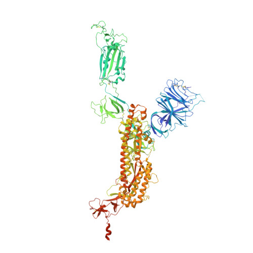
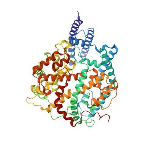
8WLO

Cryo-EM structure of SARS-CoV-2 prototype spike protein in complex with hippopotamus ACE2


Experimental Data Snapshot

  • Method: ELECTRON MICROSCOPY
  • Resolution: 2.62 Å
  • Aggregation State: PARTICLE 
  • Reconstruction Method: SINGLE PARTICLE 

wwPDB Validation   3D Report Full Report


This is version 1.2 of the entry. See complete history


Literature

Molecular basis of hippopotamus ACE2 binding to SARS-CoV-2.

Yang, R.Han, P.Han, P.Li, D.Zhao, R.Niu, S.Liu, K.Li, S.Tian, W.-.X.Gao, G.F.

(2024) J Virol 98: e0045124-e0045124

  • DOI: https://doi.org/10.1128/jvi.00451-24
  • Primary Citation of Related Structures:  
    8WLO, 8WLR

  • PubMed Abstract: 

    Severe acute respiratory syndrome coronavirus 2 (SARS-CoV-2) has a wide range of hosts, including hippopotami, which are semi-aquatic mammals and phylogenetically closely related to Cetacea. In this study, we characterized the binding properties of hippopotamus angiotensin-converting enzyme 2 (hiACE2) to the spike (S) protein receptor binding domains (RBDs) of the SARS-CoV-2 prototype (PT) and variants of concern (VOCs). Furthermore, the cryo-electron microscopy (cryo-EM) structure of the SARS-CoV-2 PT S protein complexed with hiACE2 was resolved. Structural and mutational analyses revealed that L30 and F83, which are specific to hiACE2, played a crucial role in the hiACE2/SARS-CoV-2 RBD interaction. In addition, comparative and structural analysis of ACE2 orthologs suggested that the cetaceans may have the potential to be infected by SARS-CoV-2. These results provide crucial molecular insights into the susceptibility of hippopotami to SARS-CoV-2 and suggest the potential risk of SARS-CoV-2 VOCs spillover and the necessity for surveillance. The hippopotami are the first semi-aquatic artiodactyl mammals wherein SARS-CoV-2 infection has been reported. Exploration of the invasion mechanism of SARS-CoV-2 will provide important information for the surveillance of SARS-CoV-2 in hippopotami, as well as other semi-aquatic mammals and cetaceans. Here, we found that hippopotamus ACE2 (hiACE2) could efficiently bind to the RBDs of the SARS-CoV-2 prototype (PT) and variants of concern (VOCs) and facilitate the transduction of SARS-CoV-2 PT and VOCs pseudoviruses into hiACE2-expressing cells. The cryo-EM structure of the SARS-CoV-2 PT S protein complexed with hiACE2 elucidated a few critical residues in the RBD/hiACE2 interface, especially L30 and F83 of hiACE2 which are unique to hiACE2 and contributed to the decreased binding affinity to PT RBD compared to human ACE2. Our work provides insight into cross-species transmission and highlights the necessity for monitoring host jumps and spillover events on SARS-CoV-2 in semi-aquatic/aquatic mammals.


  • Organizational Affiliation

    College of Veterinary Medicine, Shanxi Agricultural University, Jinzhong, China.


Macromolecules
Find similar proteins by:  (by identity cutoff)  |  3D Structure
Entity ID: 1
MoleculeChains Sequence LengthOrganismDetailsImage
Spike glycoprotein
A, B, C
1,217Severe acute respiratory syndrome coronavirus 2Mutation(s): 9 
Gene Names: S2
UniProt
Find proteins for P0DTC2 (Severe acute respiratory syndrome coronavirus 2)
Explore P0DTC2 
Go to UniProtKB:  P0DTC2
Entity Groups  
Sequence Clusters30% Identity50% Identity70% Identity90% Identity95% Identity100% Identity
UniProt GroupP0DTC2
Sequence Annotations
Expand
  • Reference Sequence
Find similar proteins by:  (by identity cutoff)  |  3D Structure
Entity ID: 2
MoleculeChains Sequence LengthOrganismDetailsImage
Angiotensin-converting enzymeD [auth E]597Hippopotamus amphibiusMutation(s): 0 
Entity Groups  
Sequence Clusters30% Identity50% Identity70% Identity90% Identity95% Identity100% Identity
Sequence Annotations
Expand
  • Reference Sequence
Oligosaccharides

Help

Entity ID: 3
MoleculeChains Length2D Diagram Glycosylation3D Interactions
2-acetamido-2-deoxy-beta-D-glucopyranose-(1-4)-2-acetamido-2-deoxy-beta-D-glucopyranose
E [auth D],
F,
G,
H,
I,
E [auth D],
F,
G,
H,
I,
J,
K,
L,
M,
N,
O,
P
2N-Glycosylation
Glycosylation Resources
GlyTouCan:  G42666HT
GlyCosmos:  G42666HT
GlyGen:  G42666HT
Small Molecules
Ligands 2 Unique
IDChains Name / Formula / InChI Key2D Diagram3D Interactions
NAG (Subject of Investigation/LOI)
Query on NAG

Download Ideal Coordinates CCD File 
AA [auth B]
BA [auth B]
CA [auth B]
DA [auth B]
EA [auth B]
AA [auth B],
BA [auth B],
CA [auth B],
DA [auth B],
EA [auth B],
FA [auth B],
GA [auth B],
HA [auth B],
IA [auth C],
JA [auth C],
KA [auth C],
LA [auth C],
MA [auth C],
NA [auth C],
OA [auth C],
Q [auth A],
R [auth A],
S [auth A],
T [auth A],
U [auth A],
V [auth A],
W [auth B],
X [auth B],
Y [auth B],
Z [auth B]
2-acetamido-2-deoxy-beta-D-glucopyranose
C8 H15 N O6
OVRNDRQMDRJTHS-FMDGEEDCSA-N
ZN (Subject of Investigation/LOI)
Query on ZN

Download Ideal Coordinates CCD File 
PA [auth E]ZINC ION
Zn
PTFCDOFLOPIGGS-UHFFFAOYSA-N
Experimental Data & Validation

Experimental Data

  • Method: ELECTRON MICROSCOPY
  • Resolution: 2.62 Å
  • Aggregation State: PARTICLE 
  • Reconstruction Method: SINGLE PARTICLE 

Structure Validation

View Full Validation Report


Entry History & Funding Information

Deposition Data


Funding OrganizationLocationGrant Number
Chinese Academy of SciencesChinaXDB29010202

Revision History  (Full details and data files)

  • Version 1.0: 2024-03-27
    Type: Initial release
  • Version 1.1: 2024-05-22
    Changes: Database references
  • Version 1.2: 2024-05-29
    Changes: Database references