8K7Q

Staphylococcus aureus lipase S116A inactive mutant-PSA complex


Experimental Data Snapshot

  • Method: X-RAY DIFFRACTION
  • Resolution: 2.02 Å
  • R-Value Free: 0.239 
  • R-Value Work: 0.218 
  • R-Value Observed: 0.219 

wwPDB Validation   3D Report Full Report


Ligand Structure Quality Assessment 


This is version 1.0 of the entry. See complete history


Literature

Crystal structure of Staphylococcus aureus lipase complex with unsaturated petroselinic acid.

Kitadokoro, J.Kamitani, S.Okuno, Y.Hikima, T.Yamamoto, M.Hirokawa, T.Kitadokoro, K.

(2024) FEBS Open Bio 

  • DOI: https://doi.org/10.1002/2211-5463.13808
  • Primary Citation of Related Structures:  
    8K7P, 8K7Q, 8YIB

  • PubMed Abstract: 

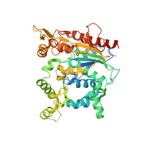
    Staphylococcus aureus produces large amounts of toxins and virulence factors. In patients with underlying diseases or compromised immune systems, this bacterium can lead to severe infections and potentially death. In this study, the crystal structure of the complex of S. aureus lipase (SAL), which is involved in the growth of this bacterium, with petroselinic acid (PSA), an inhibitor of unsaturated fatty acids, was determined by X-ray crystallography. Recently, PSA was shown to inhibit S. aureus biofilm formation and the enzymatic activity of SAL. To further characterize the inhibitory mechanism, we determined the half-inhibitory concentration of SAL by PSA and the crystal structure of the complex. The IC 50 of the inhibitory effect of PSA on SAL was 3.4 μm. SAL and PSA inhibitors were co-crystallized, and diffraction data sets were collected to 2.19 Å resolution at SPring-8 to determine the crystal structure and elucidate the detailed structural interactions. The results show that the fatty acid moiety of PSA is tightly bound to a hydrophobic pocket extending in two directions around the catalytic residue Ser116. Ser116 was also covalently bonded to the carbon of the unsaturated fatty acid moiety, and an oxyanion hole in SAL stabilized the electrons of the double bond. The difference in inhibitory activity between PSA and ester compounds revealed a structure-activity relationship between SAL and PSA. Additional research is required to further characterize the clinical potential of PSA.


  • Organizational Affiliation

    Faculty of Molecular Chemistry and Engineering, Graduate School of Science and Technology, Kyoto Institute of Technology, Japan.


Macromolecules
Find similar proteins by:  (by identity cutoff)  |  3D Structure
Entity ID: 1
MoleculeChains Sequence LengthOrganismDetailsImage
Lipase 2
A, B
408Staphylococcus aureusMutation(s): 1 
Gene Names: lipBN1321_80040
EC: 3.1.1.3
UniProt
Find proteins for A0A0U1MWF9 (Staphylococcus aureus)
Explore A0A0U1MWF9 
Go to UniProtKB:  A0A0U1MWF9
Entity Groups  
Sequence Clusters30% Identity50% Identity70% Identity90% Identity95% Identity100% Identity
UniProt GroupA0A0U1MWF9
Sequence Annotations
Expand
  • Reference Sequence
Small Molecules
Ligands 13 Unique
IDChains Name / Formula / InChI Key2D Diagram3D Interactions
4I1 (Subject of Investigation/LOI)
Query on 4I1

Download Ideal Coordinates CCD File 
BA [auth B],
H [auth A]
Petroselinic acid
C18 H34 O2
CNVZJPUDSLNTQU-SEYXRHQNSA-N
DAO
Query on DAO

Download Ideal Coordinates CCD File 
GA [auth B],
I [auth A]
LAURIC ACID
C12 H24 O2
POULHZVOKOAJMA-UHFFFAOYSA-N
OCA
Query on OCA

Download Ideal Coordinates CCD File 
AA [auth B],
D [auth A],
E [auth A],
Z [auth B]
OCTANOIC ACID (CAPRYLIC ACID)
C8 H16 O2
WWZKQHOCKIZLMA-UHFFFAOYSA-N
6NA
Query on 6NA

Download Ideal Coordinates CCD File 
HA [auth B],
J [auth A],
U [auth B],
X [auth B]
HEXANOIC ACID
C6 H12 O2
FUZZWVXGSFPDMH-UHFFFAOYSA-N
GOL
Query on GOL

Download Ideal Coordinates CCD File 
MA [auth B],
T [auth A]
GLYCEROL
C3 H8 O3
PEDCQBHIVMGVHV-UHFFFAOYSA-N
BUA
Query on BUA

Download Ideal Coordinates CCD File 
Y [auth B]butanoic acid
C4 H8 O2
FERIUCNNQQJTOY-UHFFFAOYSA-N
PPI
Query on PPI

Download Ideal Coordinates CCD File 
F [auth A]PROPANOIC ACID
C3 H6 O2
XBDQKXXYIPTUBI-UHFFFAOYSA-N
ZN (Subject of Investigation/LOI)
Query on ZN

Download Ideal Coordinates CCD File 
KA [auth B],
R [auth A]
ZINC ION
Zn
PTFCDOFLOPIGGS-UHFFFAOYSA-N
ACY
Query on ACY

Download Ideal Coordinates CCD File 
G [auth A]ACETIC ACID
C2 H4 O2
QTBSBXVTEAMEQO-UHFFFAOYSA-N
FMT
Query on FMT

Download Ideal Coordinates CCD File 
C [auth A]
CA [auth B]
DA [auth B]
EA [auth B]
FA [auth B]
C [auth A],
CA [auth B],
DA [auth B],
EA [auth B],
FA [auth B],
K [auth A],
L [auth A],
M [auth A],
N [auth A],
V [auth B],
W [auth B]
FORMIC ACID
C H2 O2
BDAGIHXWWSANSR-UHFFFAOYSA-N
CA (Subject of Investigation/LOI)
Query on CA

Download Ideal Coordinates CCD File 
LA [auth B],
S [auth A]
CALCIUM ION
Ca
BHPQYMZQTOCNFJ-UHFFFAOYSA-N
CL
Query on CL

Download Ideal Coordinates CCD File 
IA [auth B],
JA [auth B],
O [auth A],
P [auth A]
CHLORIDE ION
Cl
VEXZGXHMUGYJMC-UHFFFAOYSA-M
MG
Query on MG

Download Ideal Coordinates CCD File 
Q [auth A]MAGNESIUM ION
Mg
JLVVSXFLKOJNIY-UHFFFAOYSA-N
Experimental Data & Validation

Experimental Data

  • Method: X-RAY DIFFRACTION
  • Resolution: 2.02 Å
  • R-Value Free: 0.239 
  • R-Value Work: 0.218 
  • R-Value Observed: 0.219 
  • Space Group: P 41 2 2
Unit Cell:
Length ( Å )Angle ( ˚ )
a = 131.348α = 90
b = 131.348β = 90
c = 249.851γ = 90
Software Package:
Software NamePurpose
REFMACrefinement
XDSdata reduction
XSCALEdata scaling
PHASERphasing

Structure Validation

View Full Validation Report


Ligand Structure Quality Assessment 


Entry History & Funding Information

Deposition Data


Funding OrganizationLocationGrant Number
Not funded--

Revision History  (Full details and data files)

  • Version 1.0: 2024-06-05
    Type: Initial release