


X-ray structure of the haloalkane dehalogenase dead variant HaloTag7-D106A bound to a chloroalkane tetramethylrhodamine fluorophore ligand (CA-TMR)
Tarnawski, M., Kompa, J., Johnsson, K., Hiblot, J.To be published.
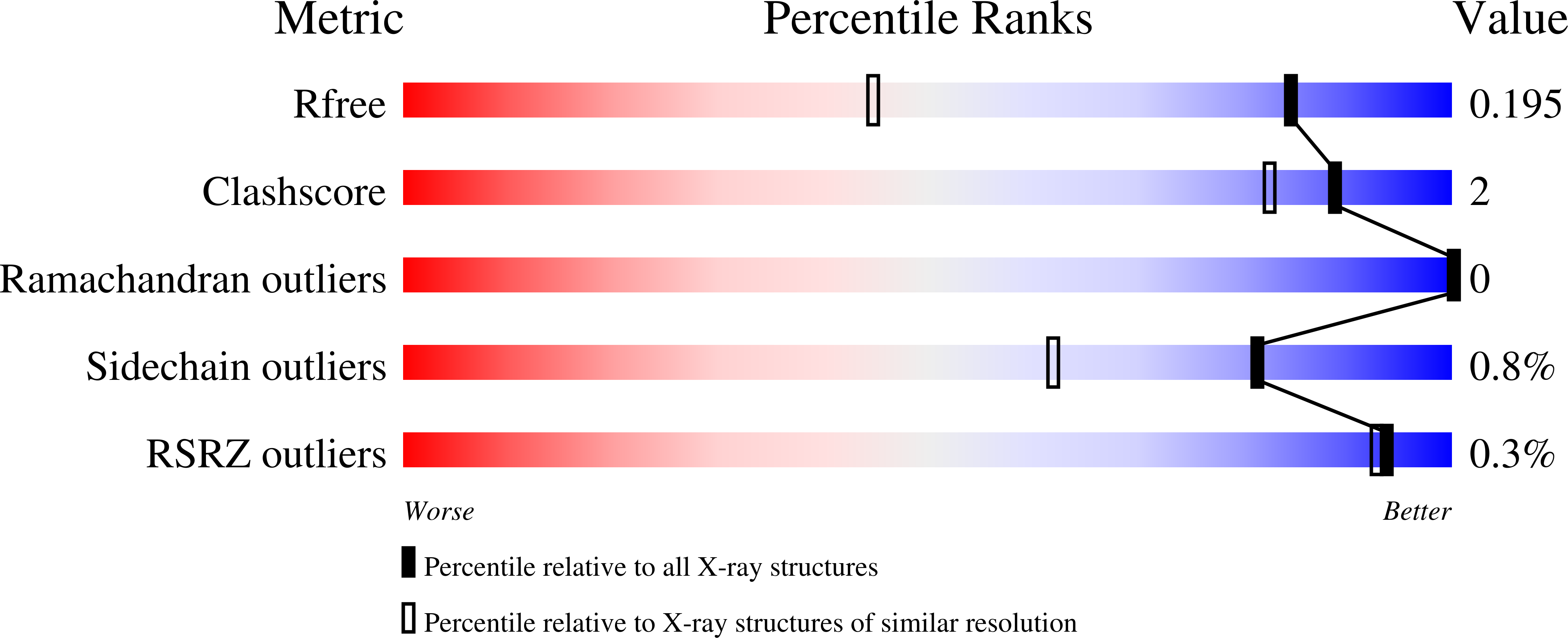
Experimental Data Snapshot
Entity ID: 1 | |||||
|---|---|---|---|---|---|
| Molecule | Chains | Sequence Length | Organism | Details | Image |
| Haloalkane dehalogenase | 293 | Rhodococcus sp. (in: high G+C Gram-positive bacteria) | Mutation(s): 22 Gene Names: dhaA EC: 3.8.1.5 | 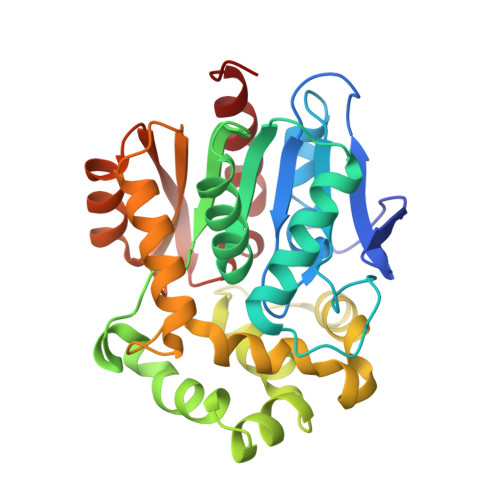 | |
UniProt | |||||
Find proteins for P0A3G3 (Rhodococcus sp) Explore P0A3G3 Go to UniProtKB: P0A3G3 | |||||
Entity Groups | |||||
| Sequence Clusters | 30% Identity50% Identity70% Identity90% Identity95% Identity100% Identity | ||||
| UniProt Group | P0A3G3 | ||||
Sequence AnnotationsExpand | |||||
| |||||
| Ligands 3 Unique | |||||
|---|---|---|---|---|---|
| ID | Chains | Name / Formula / InChI Key | 2D Diagram | 3D Interactions | |
| OEH (Subject of Investigation/LOI) Query on OEH | B [auth A] | [9-[2-carboxy-5-[2-[2-(6-chloranylhexoxy)ethoxy]ethylcarbamoyl]phenyl]-6-(dimethylamino)xanthen-3-ylidene]-dimethyl-azanium C35 H43 Cl N3 O6 YORHEDWUCXZZLS-UHFFFAOYSA-O |  | ||
| GOL Query on GOL | D [auth A] | GLYCEROL C3 H8 O3 PEDCQBHIVMGVHV-UHFFFAOYSA-N |  | ||
| CL Query on CL | C [auth A] | CHLORIDE ION Cl VEXZGXHMUGYJMC-UHFFFAOYSA-M |  | ||
| Length ( Å ) | Angle ( ˚ ) |
|---|---|
| a = 44.01 | α = 90 |
| b = 77.35 | β = 118.525 |
| c = 44.29 | γ = 90 |
| Software Name | Purpose |
|---|---|
| PHENIX | refinement |
| XDS | data reduction |
| XSCALE | data scaling |
| PHASER | phasing |
| Funding Organization | Location | Grant Number |
|---|---|---|
| Max Planck Society | Germany | -- |
RCSB PDB (citation) is hosted by
RCSB PDB is a member of the

