7YQS

Neutron structure of a L-rhamnose-alpha-1,4-D-glucuronate lyase from Fusarium oxysporum 12S, L-Rha complex


Experimental Data Snapshot

  • Method: X-RAY DIFFRACTION
  • Resolution: 1.25 Å
  • R-Value Free: 0.163 
  • R-Value Work: 0.146 
  • R-Value Observed: 0.147 

  • Method: NEUTRON DIFFRACTION
  • Resolution: 1.80 Å
  • R-Value Free: 0.191 
  • R-Value Work: 0.162 
  • R-Value Observed: 0.163 

wwPDB Validation   3D Report Full Report


This is version 1.3 of the entry. See complete history


Literature

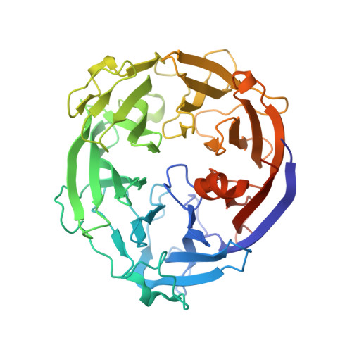
Charge neutralization and beta-elimination cleavage mechanism of family 42 L-rhamnose-alpha-1,4-D-glucuronate lyase revealed using neutron crystallography.

Yano, N.Kondo, T.Kusaka, K.Arakawa, T.Sakamoto, T.Fushinobu, S.

(2024) J Biol Chem 300: 105774-105774

  • DOI: https://doi.org/10.1016/j.jbc.2024.105774
  • Primary Citation of Related Structures:  
    7YQS, 8I4D

  • PubMed Abstract: 

    Gum arabic (GA) is widely used as an emulsion stabilizer and edible coating and consists of a complex carbohydrate moiety with a rhamnosyl-glucuronate group capping the non-reducing ends. Enzymes that can specifically cleave the glycosidic chains of GA and modify their properties are valuable for structural analysis and industrial application. Cryogenic X-ray crystal structure of GA-specific L-rhamnose-α-1,4-D-glucuronate lyase from Fusarium oxysporum (FoRham1), belonging to the polysaccharide lyase (PL) family 42, has been previously reported. To determine the specific reaction mechanism based on its hydrogen-containing enzyme structure, we performed joint X-ray/neutron crystallography of FoRham1. Large crystals were grown in the presence of L-rhamnose (a reaction product), and neutron and X-ray diffraction datasets were collected at room temperature at 1.80 and 1.25 Å resolutions, respectively. The active site contained L-rhamnose and acetate, the latter being a partial analog of glucuronate. Incomplete H/D exchange between Arg166 and acetate suggested that a strong salt-bridge interaction was maintained. Doubly deuterated His105 and deuterated Tyr150 supported the interaction between Arg166 and the acetate. The unique hydrogen-rich environment functions as a charge neutralizer for glucuronate and stabilizes the oxyanion intermediate. The NE2 atom of His85 was deprotonated and formed a hydrogen bond with the deuterated O1 hydroxy of L-rhamnose, indicating the function of His85 as the base/acid catalyst for bond cleavage via β-elimination. Asp83 functions as a pivot between the two catalytic histidine residues by bridging them. This His-His-Asp structural motif is conserved in the PL 24, 25, and 42 families.


  • Organizational Affiliation

    Structural Biology Division, Japan Synchrotron Radiation Research Institute, Hyogo, Japan. Electronic address: naomine.yano@spring8.or.jp.


Macromolecules
Find similar proteins by:  (by identity cutoff)  |  3D Structure
Entity ID: 1
MoleculeChains Sequence LengthOrganismDetailsImage
L-RHAMNOSE-ALPHA-1,4-D-GLUCURONATE LYASE443Fusarium oxysporumMutation(s): 0 
UniProt
Find proteins for A0A8D5PZ14 (Fusarium oxysporum)
Explore A0A8D5PZ14 
Go to UniProtKB:  A0A8D5PZ14
Entity Groups  
Sequence Clusters30% Identity50% Identity70% Identity90% Identity95% Identity100% Identity
UniProt GroupA0A8D5PZ14
Sequence Annotations
Expand
  • Reference Sequence
Oligosaccharides

Help

Entity ID: 2
MoleculeChains Length2D Diagram Glycosylation3D Interactions
alpha-D-mannopyranose-(1-3)-[alpha-D-mannopyranose-(1-6)]alpha-D-mannopyranose-(1-6)-[alpha-D-mannopyranose-(1-3)]beta-D-mannopyranose-(1-4)-2-acetamido-2-deoxy-beta-D-glucopyranose-(1-4)-2-acetamido-2-deoxy-beta-D-glucopyranose
B
7N-Glycosylation
Glycosylation Resources
GlyTouCan:  G55220VL
GlyCosmos:  G55220VL
GlyGen:  G55220VL
Small Molecules
Ligands 5 Unique
IDChains Name / Formula / InChI Key2D Diagram3D Interactions
RAM (Subject of Investigation/LOI)
Query on RAM

Download Ideal Coordinates CCD File 
C [auth A]alpha-L-rhamnopyranose
C6 H12 O5
SHZGCJCMOBCMKK-HGVZOGFYSA-N
TRS
Query on TRS

Download Ideal Coordinates CCD File 
E [auth A]2-AMINO-2-HYDROXYMETHYL-PROPANE-1,3-DIOL
C4 H12 N O3
LENZDBCJOHFCAS-UHFFFAOYSA-O
ACT
Query on ACT

Download Ideal Coordinates CCD File 
D [auth A]ACETATE ION
C2 H3 O2
QTBSBXVTEAMEQO-UHFFFAOYSA-M
CA
Query on CA

Download Ideal Coordinates CCD File 
F [auth A]CALCIUM ION
Ca
BHPQYMZQTOCNFJ-UHFFFAOYSA-N
NA
Query on NA

Download Ideal Coordinates CCD File 
G [auth A]SODIUM ION
Na
FKNQFGJONOIPTF-UHFFFAOYSA-N
Experimental Data & Validation

Experimental Data

  • Method: X-RAY DIFFRACTION
  • Resolution: 1.25 Å
  • R-Value Free: 0.163 
  • R-Value Work: 0.146 
  • R-Value Observed: 0.147 
  • Space Group: P 21 21 21
  • Method: NEUTRON DIFFRACTION
  • Resolution: 1.80 Å
  • R-Value Free: 0.191 
  • R-Value Work: 0.162 
  • R-Value Observed: 0.163 
  • Space Group: P 21 21 21
Unit Cell:
Length ( Å )Angle ( ˚ )
a = 57.78α = 90
b = 65.8β = 90
c = 108.95γ = 90
Software Package:
Software NamePurpose
XDSdata reduction
Aimlessdata scaling
PHENIXphasing
PHENIXrefinement

Structure Validation

View Full Validation Report


Entry History & Funding Information

Deposition Data


Funding OrganizationLocationGrant Number
Japan Society for the Promotion of Science (JSPS)Japan19H00929
Japan Society for the Promotion of Science (JSPS)Japan15H02443
Japan Society for the Promotion of Science (JSPS)Japan26660083

Revision History  (Full details and data files)

  • Version 1.0: 2023-08-09
    Type: Initial release
  • Version 1.1: 2024-02-28
    Changes: Data collection, Database references, Refinement description
  • Version 1.2: 2024-03-06
    Changes: Database references
  • Version 1.3: 2024-03-27
    Changes: Database references