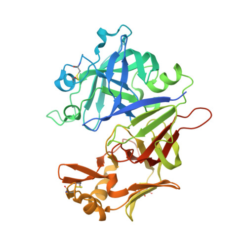
7TBC

Crystal structure of Plasmepsin X from Plasmodium falciparum in complex with WM382


Experimental Data Snapshot

  • Method: X-RAY DIFFRACTION
  • Resolution: 2.76 Å
  • R-Value Free: 0.228 
  • R-Value Work: 0.197 
  • R-Value Observed: 0.198 

wwPDB Validation   3D Report Full Report


Ligand Structure Quality Assessment 


This is version 1.2 of the entry. See complete history


Literature

Basis for drug selectivity of plasmepsin IX and X inhibition in Plasmodium falciparum and vivax.

Hodder, A.N.Christensen, J.Scally, S.Triglia, T.Ngo, A.Birkinshaw, R.W.Bailey, B.Favuzza, P.Dietrich, M.H.Tham, W.H.Czabotar, P.E.Lowes, K.Guo, Z.Murgolo, N.Lera Ruiz, M.McCauley, J.A.Sleebs, B.E.Olsen, D.Cowman, A.F.

(2022) Structure 30: 947

  • DOI: https://doi.org/10.1016/j.str.2022.03.018
  • Primary Citation of Related Structures:  
    7TBB, 7TBC, 7TBD, 7TBE

  • PubMed Abstract: 

    Plasmepsins IX (PMIX) and X (PMX) are essential aspartyl proteases for Plasmodium spp. egress, invasion, and development. WM4 and WM382 inhibit PMIX and PMX in Plasmodium falciparum and P. vivax. WM4 inhibits PMX, while WM382 is a dual inhibitor of PMIX and PMX. To understand their function, we identified protein substrates. Enzyme kinetic and structural analyses identified interactions responsible for drug specificity. PMIX and PMX have similar substrate specificity; however, there are distinct differences for peptide and protein substrates. Differences in WM4 and WM382 binding for PMIX and PMX map to variations in the S' region and engagement of the active site S3 pocket. Structures of PMX reveal interactions and mechanistic detail of drug binding important for development of clinical candidates against these targets.


  • Organizational Affiliation

    The Walter and Eliza Hall Institute of Medical Research, Parkville 3052 VIC, Australia; University of Melbourne, Melbourne, 3010 VIC, Australia.


Macromolecules
Find similar proteins by:  (by identity cutoff)  |  3D Structure
Entity ID: 1
MoleculeChains Sequence LengthOrganismDetailsImage
Plasmepsin 10377Plasmodium falciparumMutation(s): 0 
UniProt
Find proteins for Q8IAS0 (Plasmodium falciparum (isolate 3D7))
Explore Q8IAS0 
Go to UniProtKB:  Q8IAS0
Entity Groups  
Sequence Clusters30% Identity50% Identity70% Identity90% Identity95% Identity100% Identity
UniProt GroupQ8IAS0
Sequence Annotations
Expand
  • Reference Sequence
Small Molecules
Ligands 4 Unique
IDChains Name / Formula / InChI Key2D Diagram3D Interactions
I0L (Subject of Investigation/LOI)
Query on I0L

Download Ideal Coordinates CCD File 
S [auth A](4R)-4-[(2E)-4,4-diethyl-2-imino-6-oxo-1,3-diazinan-1-yl]-N-[(4S)-2,2-dimethyl-3,4-dihydro-2H-1-benzopyran-4-yl]-3,4-dihydro-2H-1-benzopyran-6-carboxamide
C29 H36 N4 O4
ZSZSSSHEMYULPX-FCHUYYIVSA-N
NAG
Query on NAG

Download Ideal Coordinates CCD File 
C [auth A]2-acetamido-2-deoxy-beta-D-glucopyranose
C8 H15 N O6
OVRNDRQMDRJTHS-FMDGEEDCSA-N
PG4
Query on PG4

Download Ideal Coordinates CCD File 
D [auth A]TETRAETHYLENE GLYCOL
C8 H18 O5
UWHCKJMYHZGTIT-UHFFFAOYSA-N
EDO
Query on EDO

Download Ideal Coordinates CCD File 
B [auth A]
E [auth A]
F [auth A]
G [auth A]
H [auth A]
B [auth A],
E [auth A],
F [auth A],
G [auth A],
H [auth A],
I [auth A],
J [auth A],
K [auth A],
L [auth A],
M [auth A],
N [auth A],
O [auth A],
P [auth A],
Q [auth A],
R [auth A]
1,2-ETHANEDIOL
C2 H6 O2
LYCAIKOWRPUZTN-UHFFFAOYSA-N
Experimental Data & Validation

Experimental Data

  • Method: X-RAY DIFFRACTION
  • Resolution: 2.76 Å
  • R-Value Free: 0.228 
  • R-Value Work: 0.197 
  • R-Value Observed: 0.198 
  • Space Group: P 32 2 1
Unit Cell:
Length ( Å )Angle ( ˚ )
a = 109.32α = 90
b = 109.32β = 90
c = 118.31γ = 120
Software Package:
Software NamePurpose
PHENIXrefinement
XDSdata reduction
Aimlessdata scaling
PHASERphasing

Structure Validation

View Full Validation Report


Ligand Structure Quality Assessment 


Entry History & Funding Information

Deposition Data


Funding OrganizationLocationGrant Number
Wellcome TrustUnited Kingdom109662/Z/15/Z, 202749/Z/16/Z

Revision History  (Full details and data files)

  • Version 1.0: 2022-05-04
    Type: Initial release
  • Version 1.1: 2022-07-20
    Changes: Database references
  • Version 1.2: 2023-10-18
    Changes: Data collection, Refinement description