7RJ0

Mouse Gamma S Crystallin L16 Octamer


Experimental Data Snapshot

  • Method: X-RAY DIFFRACTION
  • Resolution: 2.92 Å
  • R-Value Free: 0.290 
  • R-Value Work: 0.199 
  • R-Value Observed: 0.204 

wwPDB Validation   3D Report Full Report


This is version 1.3 of the entry. See complete history


Literature

Acquired Disorder and Asymmetry in a Domain-Swapped Model for gamma-Crystallin Aggregation.

Sagar, V.Wistow, G.

(2022) J Mol Biol 434: 167559-167559

  • DOI: https://doi.org/10.1016/j.jmb.2022.167559
  • Primary Citation of Related Structures:  
    7RJ0

  • PubMed Abstract: 

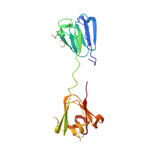
    Misfolding and aggregation of proteins occur in many pathological states. Because of the inherent disorder involved, these processes are difficult to study. We attempted to capture aggregation intermediates of γS-crystallin, a highly stable, internally symmetrical monomeric protein, by crystallization under mildly acidic and oxidizing conditions. Here we describe novel oligomerization through strained domain-swapping and partial intermolecular disulfide formation. This forms an octamer built from asymmetric tetramers, each of which comprises an asymmetric pair of twisted, domain-swapped dimers. Each tetramer shows patterns of acquired disorder among subunits, ranging from local loss of secondary structure to regions of intrinsic disorder. The octamer ring is tied together by partial intermolecular disulfide bonds, which may contribute to strain and disorder in the octamer. Oligomerization in this structure is self-limited by the distorted octamer ring. In a more heterogeneous environment, the disordered regions could serve as seeds for cascading interactions with other proteins. Indeed, solubilized protein from crystals retain many features observed in the crystal and are prone to further oligomerization and precipitation. This structure illustrates modes of loss of organized structure and aggregation that are relevant for cataract and for other disorders involving deposition of formerly well-folded proteins.


  • Organizational Affiliation

    Section on Molecular Structure and Functional Genomics, National Eye Institute, National Institutes of Health, Bethesda, MD 20892, USA.


Macromolecules
Find similar proteins by:  (by identity cutoff)  |  3D Structure
Entity ID: 1
MoleculeChains Sequence LengthOrganismDetailsImage
Gamma-crystallin SA [auth B],
B [auth A],
C,
D
177Mus musculusMutation(s): 0 
Gene Names: Crygs
UniProt
Find proteins for O35486 (Mus musculus)
Explore O35486 
Go to UniProtKB:  O35486
Entity Groups  
Sequence Clusters30% Identity50% Identity70% Identity90% Identity95% Identity100% Identity
UniProt GroupO35486
Sequence Annotations
Expand
  • Reference Sequence
Small Molecules
Modified Residues  1 Unique
IDChains TypeFormula2D DiagramParent
MSE
Query on MSE
A [auth B],
B [auth A],
C,
D
L-PEPTIDE LINKINGC5 H11 N O2 SeMET
Experimental Data & Validation

Experimental Data

  • Method: X-RAY DIFFRACTION
  • Resolution: 2.92 Å
  • R-Value Free: 0.290 
  • R-Value Work: 0.199 
  • R-Value Observed: 0.204 
  • Space Group: P 21 21 2
Unit Cell:
Length ( Å )Angle ( ˚ )
a = 61.095α = 90
b = 77.591β = 90
c = 154.052γ = 90
Software Package:
Software NamePurpose
PHENIXrefinement
PDB_EXTRACTdata extraction
HKL-2000data reduction
HKL-2000data scaling
PHENIXphasing

Structure Validation

View Full Validation Report


Entry History 

Deposition Data

  • Released Date: 2022-04-13 
  • Deposition Author(s): Sagar, V.
  • This entry supersedes: 6MYH

Revision History  (Full details and data files)

  • Version 1.0: 2022-04-13
    Type: Initial release
  • Version 1.1: 2022-04-20
    Changes: Database references
  • Version 1.2: 2023-10-18
    Changes: Data collection, Refinement description
  • Version 1.3: 2023-11-15
    Changes: Data collection