7P8E

Crystal structure of the Receiver domain of M. truncatula cytokinin receptor MtCRE1


Experimental Data Snapshot

  • Method: X-RAY DIFFRACTION
  • Resolution: 2.50 Å
  • R-Value Free: 0.221 
  • R-Value Work: 0.173 
  • R-Value Observed: 0.176 

wwPDB Validation   3D Report Full Report


This is version 1.2 of the entry. See complete history


Literature

3D Domain Swapping Dimerization of the Receiver Domain of Cytokinin Receptor CRE1 From Arabidopsis thaliana and Medicago truncatula .

Tran, L.H.Urbanowicz, A.Jasinski, M.Jaskolski, M.Ruszkowski, M.

(2021) Front Plant Sci 12: 756341-756341

  • DOI: https://doi.org/10.3389/fpls.2021.756341
  • Primary Citation of Related Structures:  
    7P8C, 7P8D, 7P8E

  • PubMed Abstract: 

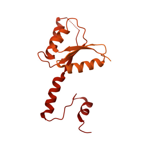
    Cytokinins are phytohormones regulating many biological processes that are vital to plants. CYTOKININ RESPONSE1 (CRE1), the main cytokinin receptor, has a modular architecture composed of a cytokinin-binding CHASE (Cyclases/Histidine kinases Associated Sensory Extracellular) domain, followed by a transmembrane fragment, an intracellular histidine kinase (HK) domain, and a receiver domain (REC). Perception of cytokinin signaling involves (i) a hormone molecule binding to the CHASE domain, (ii) CRE1 autophosphorylation at a conserved His residue in the HK domain, followed by a phosphorelay to (iii) a conserved Asp residue in the REC domain, (iv) a histidine-containing phosphotransfer protein (HPt), and (v) a response regulator (RR). This work focuses on the crystal structures of the REC domain of CRE1 from the model plant Arabidopsis thaliana and from the model legume Medicago truncatula . Both REC domains form tight 3D-domain-swapped dimers. Dimerization of the REC domain agrees with the quaternary assembly of the entire CRE1 but is incompatible with a model of its complex with HPt, suggesting that a considerable conformational change should occur to enable the signal transduction. Indeed, phosphorylation of the REC domain can change the HPt-binding properties of CRE1, as shown by functional studies.


  • Organizational Affiliation

    Institute of Bioorganic Chemistry, Polish Academy of Sciences, Poznań, Poland.


Macromolecules
Find similar proteins by:  (by identity cutoff)  |  3D Structure
Entity ID: 1
MoleculeChains Sequence LengthOrganismDetailsImage
Receiver domain of histidine kinase644Medicago truncatulaMutation(s): 0 
Gene Names: 11411168MTR_8g106150MtrunA17_Chr8g0392301
EC: 2.7.13.3
UniProt
Find proteins for G7LCC3 (Medicago truncatula)
Explore G7LCC3 
Go to UniProtKB:  G7LCC3
Entity Groups  
Sequence Clusters30% Identity50% Identity70% Identity90% Identity95% Identity100% Identity
UniProt GroupG7LCC3
Sequence Annotations
Expand
  • Reference Sequence
Small Molecules
Ligands 1 Unique
IDChains Name / Formula / InChI Key2D Diagram3D Interactions
CA (Subject of Investigation/LOI)
Query on CA

Download Ideal Coordinates CCD File 
B [auth A]CALCIUM ION
Ca
BHPQYMZQTOCNFJ-UHFFFAOYSA-N
Experimental Data & Validation

Experimental Data

  • Method: X-RAY DIFFRACTION
  • Resolution: 2.50 Å
  • R-Value Free: 0.221 
  • R-Value Work: 0.173 
  • R-Value Observed: 0.176 
  • Space Group: P 31 2 1
Unit Cell:
Length ( Å )Angle ( ˚ )
a = 60.189α = 90
b = 60.189β = 90
c = 80.41γ = 120
Software Package:
Software NamePurpose
PHENIXrefinement
XDSdata reduction
STARANISOdata scaling
PHASERphasing

Structure Validation

View Full Validation Report


Entry History & Funding Information

Deposition Data


Funding OrganizationLocationGrant Number
Polish National Science CentrePolandSONATA 2018/31/D/NZ1/03630

Revision History  (Full details and data files)

  • Version 1.0: 2021-10-20
    Type: Initial release
  • Version 1.1: 2022-02-02
    Changes: Structure summary
  • Version 1.2: 2024-01-31
    Changes: Data collection, Refinement description