7P1E

Structure of KDNase from Aspergillus Terrerus in complex with 2,3-difluoro-2-keto-3-deoxynononic acid


Experimental Data Snapshot

  • Method: X-RAY DIFFRACTION
  • Resolution: 1.53 Å
  • R-Value Free: 0.220 
  • R-Value Work: 0.186 
  • R-Value Observed: 0.187 

wwPDB Validation   3D Report Full Report


Ligand Structure Quality Assessment 


This is version 2.1 of the entry. See complete history


Literature

Kinetic and Structural Characterization of Sialidases (Kdnases) from Ascomycete Fungal Pathogens.

Nejatie, A.Steves, E.Gauthier, N.Baker, J.Nesbitt, J.McMahon, S.A.Oehler, V.Thornton, N.J.Noyovitz, B.Khazaei, K.Byers, B.W.Zandberg, W.F.Gloster, T.M.Moore, M.M.Bennet, A.J.

(2021) ACS Chem Biol 16: 2632-2640

  • DOI: https://doi.org/10.1021/acschembio.1c00666
  • Primary Citation of Related Structures:  
    7P1B, 7P1D, 7P1E, 7P1F, 7P1O, 7P1Q, 7P1R, 7P1S, 7P1U, 7P1V

  • PubMed Abstract: 

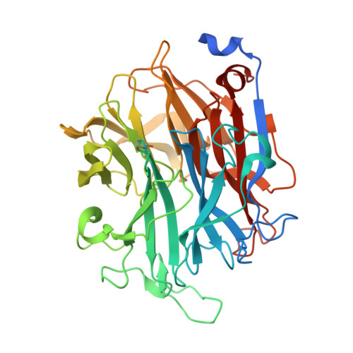
    Sialidases catalyze the release of sialic acid from the terminus of glycan chains. We previously characterized the sialidase from the opportunistic fungal pathogen, Aspergillus fumigatus, and showed that it is a Kdnase. That is, this enzyme prefers 3-deoxy-d-glycero-d-galacto-non-2-ulosonates (Kdn glycosides) as the substrate compared to N -acetylneuraminides (Neu5Ac). Here, we report characterization and crystal structures of putative sialidases from two other ascomycete fungal pathogens, Aspergillus terreus ( At S) and Trichophyton rubrum ( Tr S). Unlike A. fumigatus Kdnase ( Af S), hydrolysis with the Neu5Ac substrates was negligible for Tr S and At S; thus, Tr S and At S are selective Kdnases. The second-order rate constant for hydrolysis of aryl Kdn glycosides by At S is similar to that by Af S but 30-fold higher by Tr S. The structures of these glycoside hydrolase family 33 (GH33) enzymes in complex with a range of ligands for both At S and Tr S show subtle changes in ring conformation that mimic the Michaelis complex, transition state, and covalent intermediate formed during catalysis. In addition, they can aid identification of important residues for distinguishing between Kdn and Neu5Ac substrates. When A. fumigatus , A. terreus, and T. rubrum were grown in chemically defined media, Kdn was detected in mycelial extracts, but Neu5Ac was only observed in A. terreus or T. rubrum extracts. The C8 monosaccharide 3-deoxy-d- manno -oct-2-ulosonic acid (Kdo) was also identified in A. fumigatus and T. rubrum samples. A fluorescent Kdn probe was synthesized and revealed the localization of Af S in vesicles at the cell surface.


  • Organizational Affiliation

    Department of Chemistry, Simon Fraser University, 8888 University Drive, Burnaby V5A 1S6, British Columbia, Canada.


Macromolecules
Find similar proteins by:  (by identity cutoff)  |  3D Structure
Entity ID: 1
MoleculeChains Sequence LengthOrganismDetailsImage
Sialidase domain-containing protein410Aspergillus terreus NIH2624Mutation(s): 1 
Gene Names: ATEG_04964
UniProt
Find proteins for Q0CMX0 (Aspergillus terreus (strain NIH 2624 / FGSC A1156))
Explore Q0CMX0 
Go to UniProtKB:  Q0CMX0
Entity Groups  
Sequence Clusters30% Identity50% Identity70% Identity90% Identity95% Identity100% Identity
UniProt GroupQ0CMX0
Sequence Annotations
Expand
  • Reference Sequence
Small Molecules
Ligands 5 Unique
IDChains Name / Formula / InChI Key2D Diagram3D Interactions
K99 (Subject of Investigation/LOI)
Query on K99

Download Ideal Coordinates CCD File 
C [auth A](2R,3R,4R,5R,6S)-2,3-bis(fluoranyl)-4,5-bis(oxidanyl)-6-[(1R,2R)-1,2,3-tris(oxidanyl)propyl]oxane-2-carboxylic acid
C9 H14 F2 O8
KSUIKNBQJWNZMW-AWLRDIFTSA-N
FKD (Subject of Investigation/LOI)
Query on FKD

Download Ideal Coordinates CCD File 
B [auth A]3-deoxy-3-fluoro-D-erythro-alpha-L-manno-non-2-ulopyranosonic acid
C9 H15 F O9
KOWJBKIDVGQXJZ-QMFVTVPYSA-N
GOL
Query on GOL

Download Ideal Coordinates CCD File 
D [auth A],
E [auth A]
GLYCEROL
C3 H8 O3
PEDCQBHIVMGVHV-UHFFFAOYSA-N
CA
Query on CA

Download Ideal Coordinates CCD File 
G [auth A]CALCIUM ION
Ca
BHPQYMZQTOCNFJ-UHFFFAOYSA-N
CL
Query on CL

Download Ideal Coordinates CCD File 
F [auth A]CHLORIDE ION
Cl
VEXZGXHMUGYJMC-UHFFFAOYSA-M
Modified Residues  1 Unique
IDChains TypeFormula2D DiagramParent
SNN
Query on SNN
A
L-PEPTIDE LINKINGC4 H6 N2 O2ASN
Experimental Data & Validation

Experimental Data

  • Method: X-RAY DIFFRACTION
  • Resolution: 1.53 Å
  • R-Value Free: 0.220 
  • R-Value Work: 0.186 
  • R-Value Observed: 0.187 
  • Space Group: P 21 2 21
Unit Cell:
Length ( Å )Angle ( ˚ )
a = 61.348α = 90
b = 66.743β = 90
c = 92.635γ = 90
Software Package:
Software NamePurpose
REFMACrefinement
DIALSdata reduction
DIALSdata scaling
PHASERphasing

Structure Validation

View Full Validation Report


Ligand Structure Quality Assessment 


Entry History 

Deposition Data

Revision History  (Full details and data files)

  • Version 1.0: 2021-10-20
    Type: Initial release
  • Version 1.1: 2022-05-04
    Changes: Data collection, Database references
  • Version 2.0: 2023-11-15
    Changes: Atomic model, Data collection, Derived calculations
  • Version 2.1: 2024-01-31
    Changes: Refinement description