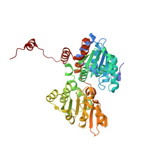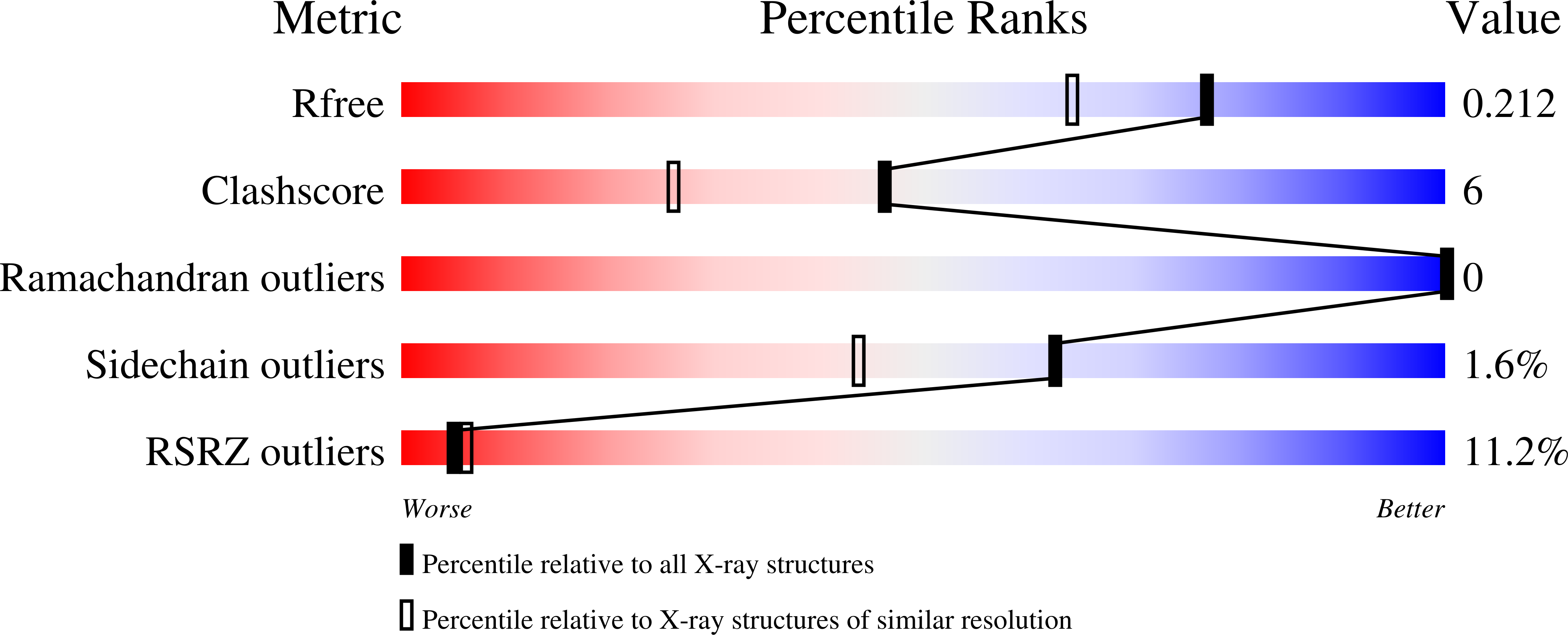
Biochemical and structural insights into an unusual, alkali-metal-independent S-adenosyl-L-homocysteine hydrolase from Synechocystis sp. PCC 6803.
Malecki, P.H., Imiolczyk, B., Barciszewski, J., Czyrko-Horczak, J., Sliwiak, J., Gawel, M., Wozniak, K., Jaskolski, M., Brzezinski, K.(2022) Acta Crystallogr D Struct Biol 78: 865-882
- PubMed: 35775986
- DOI: https://doi.org/10.1107/S2059798322005605
- Primary Citation of Related Structures:
7O5L, 7O5M, 7ZD7, 7ZD8, 7ZD9 - PubMed Abstract:
The mesophilic cyanobacterium Synechocystis sp. PCC 6803 encodes an S-adenosyl-L-homocysteine hydrolase (SAHase) of archaeal origin in its genome. SAHases are essential enzymes involved in the regulation of cellular S-adenosyl-L-methionine (SAM)-dependent methylation reactions. They are usually active as homotetramers or, less commonly, as homodimers. A SAHase subunit is composed of two major domains: a cofactor (NAD + )-binding domain and a substrate (S-adenosyl-L-homocysteine)-binding domain. These are connected by a hinge element that is also a coordination site for an alkali-metal cation that influences domain movement during the catalytic cycle. Typically, the highest activity and strongest substrate binding of bacterial SAHases are observed in the presence of K + ions. The SAHase from Synechocystis (SynSAHase) is an exception in this respect. Enzymatic and isothermal titration calorimetry studies demonstrated that in contrast to K + -dependent SAHases, the activity and ligand binding of SynSAHase are not affected by the presence of any particular alkali ion. Moreover, in contrast to other SAHases, the cyanobacterial enzyme is in an equilibrium of two distinct oligomeric states corresponding to its dimeric and tetrameric forms in solution. To explain these phenomena, crystal structures of SynSAHase were determined for the enzyme crystallized in the presence of adenosine (a reaction byproduct or substrate) and sodium or rubidium cations. The structural data confirm that while SynSAHase shares common structural features with other SAHases, no alkali metal is coordinated by the cyanobacterial enzyme as a result of a different organization of the macromolecular environment of the site that is normally supposed to coordinate the metal cation. This inspired the generation of SynSAHase mutants that bind alkali-metal cations analogously to K + -dependent SAHases, as confirmed by crystallographic studies. Structural comparisons of the crystal structure of SynSAHase with other experimental models of SAHases suggest a possible explanation for the occurrence of the cyanobacterial enzyme in the tetrameric state. On the other hand, the reason for the existence of SynSAHase in the dimeric state in solution remains elusive.
Organizational Affiliation:
Department of Structural Biology of Prokaryotic Organisms, Institute of Bioorganic Chemistry, Polish Academy of Sciences, Noskowskiego 12/14, 61-704 Poznan, Poland.