7LYV

Crystal structure of the HCMV pentamer-specific antibody 1-103


Experimental Data Snapshot

  • Method: X-RAY DIFFRACTION
  • Resolution: 1.90 Å
  • R-Value Free: 0.217 
  • R-Value Work: 0.191 

wwPDB Validation   3D Report Full Report


This is version 1.2 of the entry. See complete history


Literature

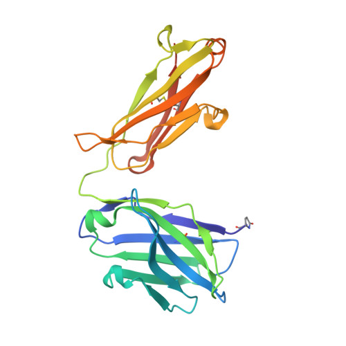
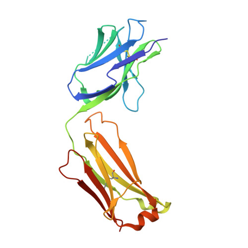
Structural basis for HCMV Pentamer recognition by neuropilin 2 and neutralizing antibodies.

Wrapp, D.Ye, X.Ku, Z.Su, H.Jones, H.G.Wang, N.Mishra, A.K.Freed, D.C.Li, F.Tang, A.Li, L.Jaijyan, D.K.Zhu, H.Wang, D.Fu, T.M.Zhang, N.An, Z.McLellan, J.S.

(2022) Sci Adv 8: eabm2546-eabm2546

  • DOI: https://doi.org/10.1126/sciadv.abm2546
  • Primary Citation of Related Structures:  
    7KBA, 7KBB, 7LYV, 7LYW, 7M1C, 7M22, 7M30

  • PubMed Abstract: 

    Human cytomegalovirus (HCMV) encodes multiple surface glycoprotein complexes to infect a variety of cell types. The HCMV Pentamer, composed of gH, gL, UL128, UL130, and UL131A, enhances entry into epithelial, endothelial, and myeloid cells by interacting with the cell surface receptor neuropilin 2 (NRP2). Despite the critical nature of this interaction, the molecular determinants that govern NRP2 recognition remain unclear. Here, we describe the cryo-EM structure of NRP2 bound to Pentamer. The high-affinity interaction between these proteins is calcium dependent and differs from the canonical carboxyl-terminal arginine (CendR) binding that NRP2 typically uses. We also determine the structures of four neutralizing human antibodies bound to the HCMV Pentamer to define susceptible epitopes. Two of these antibodies compete with NRP2 binding, but the two most potent antibodies recognize a previously unidentified epitope that does not overlap the NRP2-binding site. Collectively, these findings provide a structural basis for HCMV tropism and antibody-mediated neutralization.


  • Organizational Affiliation

    Department of Molecular Biosciences, The University of Texas at Austin, Austin, TX 78712, USA.


Macromolecules
Find similar proteins by:  (by identity cutoff)  |  3D Structure
Entity ID: 1
MoleculeChains Sequence LengthOrganismDetailsImage
1-103 Fab Heavy ChainA [auth H],
C,
E [auth A],
G [auth E]
232Homo sapiensMutation(s): 0 
Entity Groups  
Sequence Clusters30% Identity50% Identity70% Identity90% Identity95% Identity100% Identity
Sequence Annotations
Expand
  • Reference Sequence
Find similar proteins by:  (by identity cutoff)  |  3D Structure
Entity ID: 2
MoleculeChains Sequence LengthOrganismDetailsImage
1-103 Fab Light ChainB [auth L],
D,
F [auth B],
H [auth F]
210Homo sapiensMutation(s): 0 
Entity Groups  
Sequence Clusters30% Identity50% Identity70% Identity90% Identity95% Identity100% Identity
Sequence Annotations
Expand
  • Reference Sequence
Small Molecules
Modified Residues  1 Unique
IDChains TypeFormula2D DiagramParent
PCA
Query on PCA
A [auth H],
C,
E [auth A],
G [auth E]
L-PEPTIDE LINKINGC5 H7 N O3GLN
Experimental Data & Validation

Experimental Data

  • Method: X-RAY DIFFRACTION
  • Resolution: 1.90 Å
  • R-Value Free: 0.217 
  • R-Value Work: 0.191 
  • Space Group: P 1 2 1
Unit Cell:
Length ( Å )Angle ( ˚ )
a = 95.389α = 90
b = 105.24β = 91.37
c = 102.73γ = 90
Software Package:
Software NamePurpose
Aimlessdata scaling
PHENIXrefinement
PDB_EXTRACTdata extraction
MOSFLMdata reduction
PHASERphasing

Structure Validation

View Full Validation Report


Entry History 

Deposition Data

Revision History  (Full details and data files)

  • Version 1.0: 2021-08-11
    Type: Initial release
  • Version 1.1: 2022-03-23
    Changes: Database references
  • Version 1.2: 2023-10-18
    Changes: Data collection, Refinement description