7LQE

Cryo-EM of 1-protofilament of the KFE8 thinner nanotube


Experimental Data Snapshot

  • Method: ELECTRON MICROSCOPY
  • Resolution: 3.40 Å
  • Aggregation State: FILAMENT 
  • Reconstruction Method: HELICAL 

wwPDB Validation   3D Report Full Report


This is version 2.0 of the entry. See complete history


Literature

Deterministic chaos in the self-assembly of beta sheet nanotubes from an amphipathic oligopeptide.

Wang, F.Gnewou, O.Wang, S.Osinski, T.Zuo, X.Egelman, E.H.Conticello, V.P.

(2021) Matter 4: 3217-3231

  • DOI: https://doi.org/10.1016/j.matt.2021.06.037
  • Primary Citation of Related Structures:  
    7LQE, 7LQF, 7LQG, 7LQH, 7LQI

  • PubMed Abstract: 

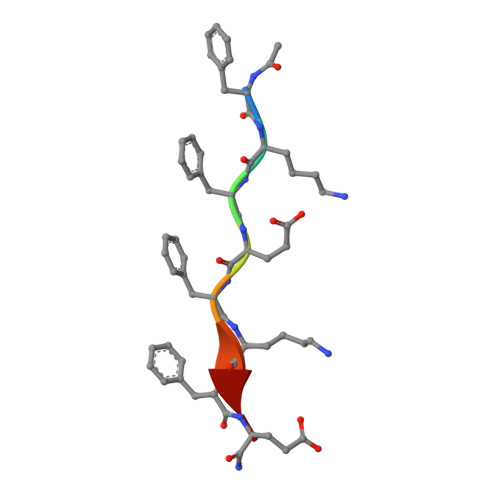
    The self-assembly of designed peptides into filaments and other higher-order structures has been the focus of intense interest because of the potential for creating new biomaterials and biomedical devices. These peptide assemblies have also been used as models for understanding biological processes, such as the pathological formation of amyloid. We investigate the assembly of an octapeptide sequence, Ac-FKFEFKFE-NH 2 , motivated by prior studies that demonstrated that this amphipathic β strand peptide self-assembled into fibrils and biocompatible hydrogels. Using high-resolution cryoelectron microscopy (cryo-EM), we are able to determine the atomic structure for two different coexisting forms of the fibrils, containing four and five β sandwich protofilaments, respectively. Surprisingly, the inner walls in both forms are parallel β sheets, while the outer walls are antiparallel β sheets. Our results demonstrate the chaotic nature of peptide self-assembly and illustrate the importance of cryo-EM structural analysis to understand the complex phase behavior of these materials at near-atomic resolution.


  • Organizational Affiliation

    Department of Biochemistry and Molecular Genetics, University of Virginia, Charlottesville, VA 22908, USA.


Macromolecules

Find similar proteins by:  Sequence   |   3D Structure  

Entity ID: 1
MoleculeChains Sequence LengthOrganismDetailsImage
KFE8 peptide8synthetic constructMutation(s): 0 
Sequence Annotations
Expand
  • Reference Sequence
Small Molecules
Modified Residues  2 Unique
IDChains TypeFormula2D DiagramParent
5CR
Query on 5CR
A
AA [auth l]
AB [auth e]
B [auth J]
BA [auth m]
A,
AA [auth l],
AB [auth e],
B [auth J],
BA [auth m],
BB [auth 5],
C [auth K],
CA [auth H],
CB [auth 6],
D [auth L],
DA [auth n],
DB [auth 7],
E [auth B],
EA [auth o],
EB [auth f],
F [auth M],
FA [auth p],
FB [auth 8],
G [auth N],
GA [auth I],
GB [auth 9],
H [auth O],
HA [auth q],
HB [auth AA],
I [auth C],
IA [auth r],
IB [auth g],
J [auth P],
JA [auth s],
JB [auth BA],
K [auth Q],
KA [auth a],
KB [auth CA],
L [auth R],
LA [auth t],
LB [auth DA],
M [auth D],
MA [auth u],
MB [auth h],
N [auth S],
NA [auth v],
NB [auth EA],
O [auth T],
OA [auth b],
OB [auth FA],
P [auth U],
PA [auth w],
PB [auth GA],
Q [auth E],
QA [auth x],
QB [auth i],
R [auth V],
RA [auth y],
RB [auth HA],
S [auth W],
SA [auth c],
SB [auth IA],
T [auth X],
TA [auth z],
TB [auth JA],
U [auth F],
UA [auth 0],
V [auth Y],
VA [auth 1],
W [auth Z],
WA [auth d],
X [auth j],
XA [auth 2],
Y [auth G],
YA [auth 3],
Z [auth k],
ZA [auth 4]
L-PEPTIDE LINKINGC11 H13 N O3PHE
GMA
Query on GMA
A
AA [auth l]
AB [auth e]
B [auth J]
BA [auth m]
A,
AA [auth l],
AB [auth e],
B [auth J],
BA [auth m],
BB [auth 5],
C [auth K],
CA [auth H],
CB [auth 6],
D [auth L],
DA [auth n],
DB [auth 7],
E [auth B],
EA [auth o],
EB [auth f],
F [auth M],
FA [auth p],
FB [auth 8],
G [auth N],
GA [auth I],
GB [auth 9],
H [auth O],
HA [auth q],
HB [auth AA],
I [auth C],
IA [auth r],
IB [auth g],
J [auth P],
JA [auth s],
JB [auth BA],
K [auth Q],
KA [auth a],
KB [auth CA],
L [auth R],
LA [auth t],
LB [auth DA],
M [auth D],
MA [auth u],
MB [auth h],
N [auth S],
NA [auth v],
NB [auth EA],
O [auth T],
OA [auth b],
OB [auth FA],
P [auth U],
PA [auth w],
PB [auth GA],
Q [auth E],
QA [auth x],
QB [auth i],
R [auth V],
RA [auth y],
RB [auth HA],
S [auth W],
SA [auth c],
SB [auth IA],
T [auth X],
TA [auth z],
TB [auth JA],
U [auth F],
UA [auth 0],
V [auth Y],
VA [auth 1],
W [auth Z],
WA [auth d],
X [auth j],
XA [auth 2],
Y [auth G],
YA [auth 3],
Z [auth k],
ZA [auth 4]
L-PEPTIDE LINKINGC5 H10 N2 O3GLU
Experimental Data & Validation

Experimental Data

  • Method: ELECTRON MICROSCOPY
  • Resolution: 3.40 Å
  • Aggregation State: FILAMENT 
  • Reconstruction Method: HELICAL 

Structure Validation

View Full Validation Report


Entry History & Funding Information

Deposition Data


Funding OrganizationLocationGrant Number
National Science Foundation (NSF, United States)United StatesNSF-DMR-1533958

Revision History  (Full details and data files)

  • Version 1.0: 2021-06-30
    Type: Initial release
  • Version 1.1: 2021-08-11
    Changes: Database references
  • Version 1.2: 2021-10-27
    Changes: Database references
  • Version 2.0: 2023-11-15
    Changes: Atomic model, Data collection