7KZD

Crystal structure of KabA from Bacillus cereus UW85 in complex with the reduced internal aldimine and with bound Glutarate


Experimental Data Snapshot

  • Method: X-RAY DIFFRACTION
  • Resolution: 1.90 Å
  • R-Value Free: 0.249 
  • R-Value Work: 0.207 
  • R-Value Observed: 0.209 

wwPDB Validation   3D Report Full Report


Ligand Structure Quality Assessment 


This is version 1.3 of the entry. See complete history


Literature

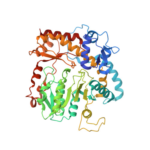
Snapshots along the catalytic path of KabA, a PLP-dependent aminotransferase required for kanosamine biosynthesis in Bacillus cereus UW85.

Prasertanan, T.Palmer, D.R.J.Sanders, D.A.R.

(2021) J Struct Biol 213: 107744-107744

  • DOI: https://doi.org/10.1016/j.jsb.2021.107744
  • Primary Citation of Related Structures:  
    7KZ3, 7KZ5, 7KZ6, 7KZD

  • PubMed Abstract: 

    Kanosamine is an antibiotic and antifungal monosaccharide. The kanosamine biosynthetic pathway from glucose 6-phosphate in Bacillus cereus UW85 was recently reported, and the functions of each of the three enzymes in the pathway, KabA, KabB and KabC, were demonstrated. KabA, a member of a subclass of the VI β family of PLP-dependent aminotransferases, catalyzes the second step in the pathway, generating kanosamine 6-phosphate (K6P) using l-glutamate as the amino-donor. KabA catalysis was shown to be extremely efficient, with a second-order rate constant with respect to K6P transamination of over 10 7 M -1 s -1 . Here we report the high-resolution structure of KabA in both the PLP- and PMP-bound forms. In addition, co-crystallization with K6P allowed the structure of KabA in complex with the covalent PLP-K6P adduct to be solved. Co-crystallization or soaking with glutamate or 2-oxoglutarate did not result in crystals with either substrate/product. Reduction of the PLP-KabA complex with sodium cyanoborohydride gave an inactivated enzyme, and crystals of the reduced KabA were soaked with the l-glutamate analog glutarate to mimic the KabA-PLP-l-glutamate complex. Together these four structures give a complete picture of how the active site of KabA recognizes substrates for each half-reaction. The KabA structure is discussed in the context of homologous aminotransferases.


  • Organizational Affiliation

    Department of Chemistry, University of Saskatchewan, Saskatoon, SK S7N 5C9, Canada.


Macromolecules
Find similar proteins by:  (by identity cutoff)  |  3D Structure
Entity ID: 1
MoleculeChains Sequence LengthOrganismDetailsImage
Aminotransferase class I/II-fold pyridoxal phosphate-dependent enzyme
A, B, C, D, E
A, B, C, D, E, F, G, H
445Bacillus cereusMutation(s): 0 
Gene Names: kabAGE376_30835
EC: 2.6.1.1
UniProt
Find proteins for C0JRF5 (Bacillus cereus)
Explore C0JRF5 
Go to UniProtKB:  C0JRF5
Entity Groups  
Sequence Clusters30% Identity50% Identity70% Identity90% Identity95% Identity100% Identity
UniProt GroupC0JRF5
Sequence Annotations
Expand
  • Reference Sequence
Small Molecules
Ligands 3 Unique
IDChains Name / Formula / InChI Key2D Diagram3D Interactions
PLR (Subject of Investigation/LOI)
Query on PLR

Download Ideal Coordinates CCD File 
DA [auth C]
DB [auth G]
I [auth A]
KA [auth D]
KB [auth H]
DA [auth C],
DB [auth G],
I [auth A],
KA [auth D],
KB [auth H],
Q [auth B],
QA [auth E],
YA [auth F]
(5-HYDROXY-4,6-DIMETHYLPYRIDIN-3-YL)METHYL DIHYDROGEN PHOSPHATE
C8 H12 N O5 P
RBCOYOYDYNXAFA-UHFFFAOYSA-N
GUA (Subject of Investigation/LOI)
Query on GUA

Download Ideal Coordinates CCD File 
CA [auth C],
JA [auth D],
RA [auth E],
ZA [auth F]
GLUTARIC ACID
C5 H8 O4
JFCQEDHGNNZCLN-UHFFFAOYSA-N
EDO
Query on EDO

Download Ideal Coordinates CCD File 
AA [auth B]
AB [auth F]
BA [auth B]
BB [auth F]
CB [auth F]
AA [auth B],
AB [auth F],
BA [auth B],
BB [auth F],
CB [auth F],
EA [auth C],
EB [auth G],
FA [auth C],
FB [auth G],
GA [auth C],
GB [auth G],
HA [auth C],
HB [auth G],
IA [auth C],
IB [auth G],
J [auth A],
JB [auth G],
K [auth A],
L [auth A],
LA [auth D],
LB [auth H],
M [auth A],
MA [auth D],
MB [auth H],
N [auth A],
NA [auth D],
NB [auth H],
O [auth A],
OA [auth D],
P [auth A],
PA [auth D],
R [auth B],
S [auth B],
SA [auth E],
T [auth B],
TA [auth E],
U [auth B],
UA [auth E],
V [auth B],
VA [auth E],
W [auth B],
WA [auth E],
X [auth B],
XA [auth E],
Y [auth B],
Z [auth B]
1,2-ETHANEDIOL
C2 H6 O2
LYCAIKOWRPUZTN-UHFFFAOYSA-N
Experimental Data & Validation

Experimental Data

  • Method: X-RAY DIFFRACTION
  • Resolution: 1.90 Å
  • R-Value Free: 0.249 
  • R-Value Work: 0.207 
  • R-Value Observed: 0.209 
  • Space Group: P 1
Unit Cell:
Length ( Å )Angle ( ˚ )
a = 92.94α = 108.84
b = 93.58β = 92.33
c = 106.89γ = 90.08
Software Package:
Software NamePurpose
PHENIXrefinement
PDB_EXTRACTdata extraction
MOLREPphasing
AutoProcessdata reduction
AutoProcessdata scaling

Structure Validation

View Full Validation Report


Ligand Structure Quality Assessment 


Entry History & Funding Information

Deposition Data


Funding OrganizationLocationGrant Number
Natural Sciences and Engineering Research Council (NSERC, Canada)Canada--

Revision History  (Full details and data files)

  • Version 1.0: 2021-05-26
    Type: Initial release
  • Version 1.1: 2021-06-02
    Changes: Database references
  • Version 1.2: 2023-10-18
    Changes: Data collection, Database references, Refinement description
  • Version 1.3: 2024-03-13
    Changes: Source and taxonomy