7CEA

Crystal structure of HUTS-4 Fv-clasp fragment


Experimental Data Snapshot

  • Method: X-RAY DIFFRACTION
  • Resolution: 2.55 Å
  • R-Value Free: 0.266 
  • R-Value Work: 0.252 
  • R-Value Observed: 0.253 

wwPDB Validation   3D Report Full Report


This is version 1.3 of the entry. See complete history


Literature

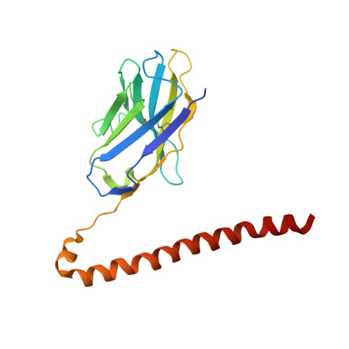
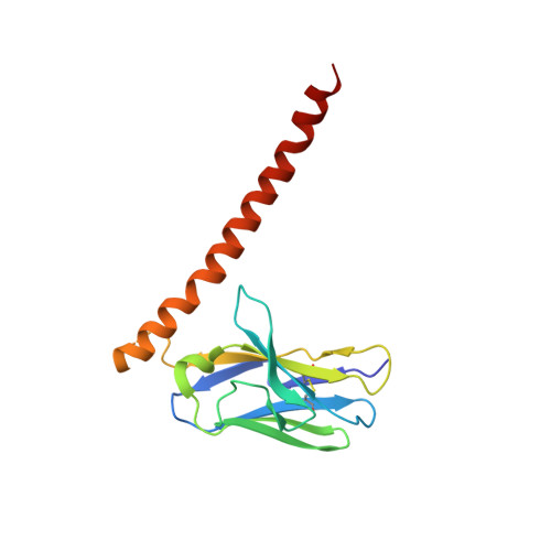
Structural mechanism of laminin recognition by integrin.

Arimori, T.Miyazaki, N.Mihara, E.Takizawa, M.Taniguchi, Y.Cabanas, C.Sekiguchi, K.Takagi, J.

(2021) Nat Commun 12: 4012-4012

  • DOI: https://doi.org/10.1038/s41467-021-24184-8
  • Primary Citation of Related Structures:  
    7CEA, 7CEB, 7CEC

  • PubMed Abstract: 

    Recognition of laminin by integrin receptors is central to the epithelial cell adhesion to basement membrane, but the structural background of this molecular interaction remained elusive. Here, we report the structures of the prototypic laminin receptor α6β1 integrin alone and in complex with three-chain laminin-511 fragment determined via crystallography and cryo-electron microscopy, respectively. The laminin-integrin interface is made up of several binding sites located on all five subunits, with the laminin γ1 chain C-terminal portion providing focal interaction using two carboxylate anchor points to bridge metal-ion dependent adhesion site of integrin β1 subunit and Asn189 of integrin α6 subunit. Laminin α5 chain also contributes to the affinity and specificity by making electrostatic interactions with large surface on the β-propeller domain of α6, part of which comprises an alternatively spliced X1 region. The propeller sheet corresponding to this region shows unusually high mobility, suggesting its unique role in ligand capture.


  • Organizational Affiliation

    Laboratory for Protein Synthesis and Expression, Institute for Protein Research, Osaka University, Suita, Osaka, Japan.


Macromolecules
Find similar proteins by:  (by identity cutoff)  |  3D Structure
Entity ID: 1
MoleculeChains Sequence LengthOrganismDetailsImage
HUTS-4 VH(S112C)-SARAH174Mus musculusHomo sapiens
This entity is chimeric
Mutation(s): 0 
UniProt & NIH Common Fund Data Resources
Find proteins for Q13043 (Homo sapiens)
Explore Q13043 
Go to UniProtKB:  Q13043
PHAROS:  Q13043
GTEx:  ENSG00000101109 
Entity Groups  
Sequence Clusters30% Identity50% Identity70% Identity90% Identity95% Identity100% Identity
UniProt GroupQ13043
Sequence Annotations
Expand
  • Reference Sequence
Find similar proteins by:  (by identity cutoff)  |  3D Structure
Entity ID: 2
MoleculeChains Sequence LengthOrganismDetailsImage
HUTS-4 VL(C87Y)-SARAH(S37C)159Mus musculusHomo sapiens
This entity is chimeric
Mutation(s): 0 
UniProt & NIH Common Fund Data Resources
Find proteins for Q13043 (Homo sapiens)
Explore Q13043 
Go to UniProtKB:  Q13043
PHAROS:  Q13043
GTEx:  ENSG00000101109 
Entity Groups  
Sequence Clusters30% Identity50% Identity70% Identity90% Identity95% Identity100% Identity
UniProt GroupQ13043
Sequence Annotations
Expand
  • Reference Sequence
Experimental Data & Validation

Experimental Data

  • Method: X-RAY DIFFRACTION
  • Resolution: 2.55 Å
  • R-Value Free: 0.266 
  • R-Value Work: 0.252 
  • R-Value Observed: 0.253 
  • Space Group: P 32 2 1
Unit Cell:
Length ( Å )Angle ( ˚ )
a = 88.367α = 90
b = 88.367β = 90
c = 78.117γ = 120
Software Package:
Software NamePurpose
PHENIXrefinement
XDSdata reduction
XDSdata scaling
PHASERphasing

Structure Validation

View Full Validation Report


Entry History & Funding Information

Deposition Data


Funding OrganizationLocationGrant Number
Japan Society for the Promotion of Science (JSPS)Japan18H02389
Japan Agency for Medical Research and Development (AMED)Japan19am0101075

Revision History  (Full details and data files)

  • Version 1.0: 2021-06-23
    Type: Initial release
  • Version 1.1: 2021-07-07
    Changes: Database references
  • Version 1.2: 2021-07-14
    Changes: Database references
  • Version 1.3: 2023-11-29
    Changes: Data collection, Database references, Refinement description