6Y98

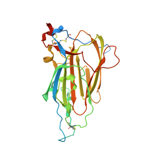
Crystal Structure of subtype-switched Epithelial Adhesin 9 to 1 A domain (Epa9-CBL2Epa1) from Candida glabrata in complex with beta-lactose


Experimental Data Snapshot

  • Method: X-RAY DIFFRACTION
  • Resolution: 2.80 Å
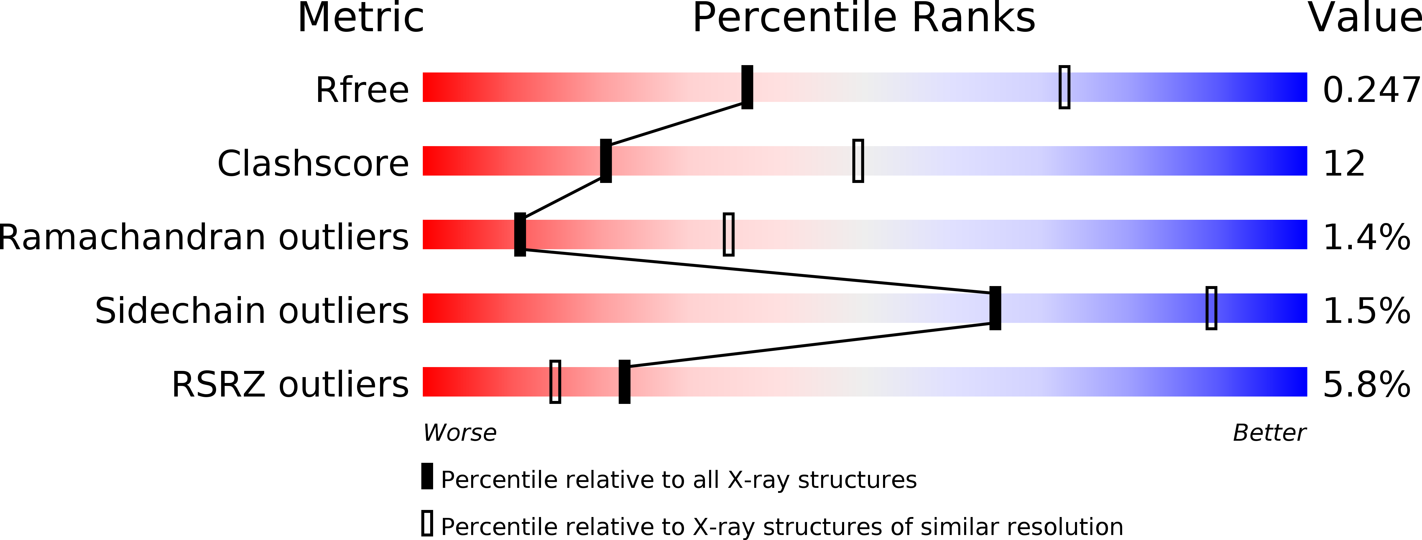
  • R-Value Free: 0.247 
  • R-Value Work: 0.204 
  • R-Value Observed: 0.206 

wwPDB Validation   3D Report Full Report


This is version 2.2 of the entry. See complete history


Literature

Functional reprogramming ofCandida glabrataepithelial adhesins: the role of conserved and variable structural motifs in ligand binding.

Hoffmann, D.Diderrich, R.Reithofer, V.Friederichs, S.Kock, M.Essen, L.O.Mosch, H.U.

(2020) J Biol Chem 295: 12512-12524

  • DOI: https://doi.org/10.1074/jbc.RA120.013968
  • Primary Citation of Related Structures:  
    6Y98, 6Y9J

  • PubMed Abstract: 

    For host-cell interaction, the human fungal pathogen Candida glabrata harbors a large family of more than 20 cell wall-attached epithelial adhesins (Epas). Epa family members are lectins with binding pockets containing several conserved and variable structural hot spots, which were implicated in mediating functional diversity. In this study, we have performed an elaborate structure-based mutational analysis of numerous Epa paralogs to generally determine the role of diverse structural hot spots in conferring host cell binding and ligand binding specificity. Our study reveals that several conserved structural motifs contribute to efficient host cell binding. Moreover, our directed motif exchange experiments reveal that the variable loop CBL2 is key for programming ligand binding specificity, albeit with limited predictability. In contrast, we find that the variable loop L1 affects host cell binding without significantly influencing the specificity of ligand binding. Our data strongly suggest that variation of numerous structural hot spots in the ligand binding pocket of Epa proteins is a main driver of their functional diversification and evolution.


  • Organizational Affiliation

    Department of Genetics, Philipps-Universität, Marburg, Germany.


Macromolecules
Find similar proteins by:  (by identity cutoff)  |  3D Structure
Entity ID: 1
MoleculeChains Sequence LengthOrganismDetailsImage
PA14 domain-containing protein290Nakaseomyces glabratus CBS 138Mutation(s): 0 
Gene Names: EPA9CAGL0A01366g
UniProt
Find proteins for B4UMX2 (Candida glabrata (strain ATCC 2001 / BCRC 20586 / JCM 3761 / NBRC 0622 / NRRL Y-65 / CBS 138))
Explore B4UMX2 
Go to UniProtKB:  B4UMX2
Entity Groups  
Sequence Clusters30% Identity50% Identity70% Identity90% Identity95% Identity100% Identity
UniProt GroupB4UMX2
Sequence Annotations
Expand
  • Reference Sequence
Oligosaccharides

Help

Entity ID: 2
MoleculeChains Length2D Diagram Glycosylation3D Interactions
beta-D-galactopyranose-(1-4)-beta-D-glucopyranose
B
2N/A
Glycosylation Resources
GlyTouCan:  G84224TW
GlyCosmos:  G84224TW
GlyGen:  G84224TW
Small Molecules
Ligands 1 Unique
IDChains Name / Formula / InChI Key2D Diagram3D Interactions
CA
Query on CA

Download Ideal Coordinates CCD File 
C [auth A]CALCIUM ION
Ca
BHPQYMZQTOCNFJ-UHFFFAOYSA-N
Biologically Interesting Molecules (External Reference) 1 Unique
Experimental Data & Validation

Experimental Data

  • Method: X-RAY DIFFRACTION
  • Resolution: 2.80 Å
  • R-Value Free: 0.247 
  • R-Value Work: 0.204 
  • R-Value Observed: 0.206 
  • Space Group: P 31 2 1
Unit Cell:
Length ( Å )Angle ( ˚ )
a = 65.289α = 90
b = 65.289β = 90
c = 121.877γ = 120
Software Package:
Software NamePurpose
PHENIXrefinement
Aimlessdata scaling
MOLREPphasing
PDB_EXTRACTdata extraction
XDSdata reduction

Structure Validation

View Full Validation Report


Entry History & Funding Information

Deposition Data


Funding OrganizationLocationGrant Number
German Research Foundation (DFG)Germany--

Revision History  (Full details and data files)

  • Version 1.0: 2020-07-22
    Type: Initial release
  • Version 2.0: 2020-07-29
    Type: Remediation
    Reason: Carbohydrate remediation
    Changes: Atomic model, Author supporting evidence, Data collection, Database references, Derived calculations, Non-polymer description, Structure summary
  • Version 2.1: 2020-09-09
    Changes: Database references
  • Version 2.2: 2024-01-24
    Changes: Data collection, Database references, Refinement description