6Y4L

Crystal structure of human ER membrane protein complex subunits EMC2 and EMC9


Experimental Data Snapshot

  • Method: X-RAY DIFFRACTION
  • Resolution: 2.20 Å
  • R-Value Free: 0.250 
  • R-Value Work: 0.203 
  • R-Value Observed: 0.206 

wwPDB Validation   3D Report Full Report


This is version 1.1 of the entry. See complete history

Re-refinement Note

A newer entry is available that reflects an alternative modeling of the original data: 6Z3W


Literature

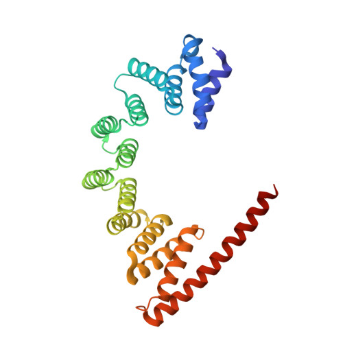
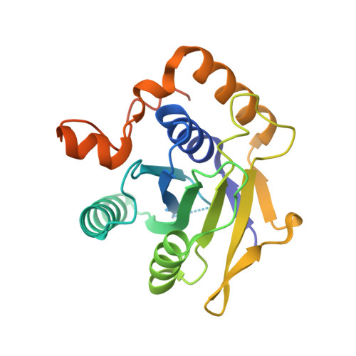
The architecture of EMC reveals a path for membrane protein insertion.

O'Donnell, J.P.Phillips, B.P.Yagita, Y.Juszkiewicz, S.Wagner, A.Malinverni, D.Keenan, R.J.Miller, E.A.Hegde, R.S.

(2020) Elife 9

  • DOI: https://doi.org/10.7554/eLife.57887
  • Primary Citation of Related Structures:  
    6Y4L, 6Z3W

  • PubMed Abstract: 

    Approximately 25% of eukaryotic genes code for integral membrane proteins that are assembled at the endoplasmic reticulum. An abundant and widely conserved multi-protein complex termed EMC has been implicated in membrane protein biogenesis, but its mechanism of action is poorly understood. Here, we define the composition and architecture of human EMC using biochemical assays, crystallography of individual subunits, site-specific photocrosslinking, and cryo-EM reconstruction. Our results suggest that EMC's cytosolic domain contains a large, moderately hydrophobic vestibule that can bind a substrate's transmembrane domain (TMD). The cytosolic vestibule leads into a lumenally-sealed, lipid-exposed intramembrane groove large enough to accommodate a single substrate TMD. A gap between the cytosolic vestibule and intramembrane groove provides a potential path for substrate egress from EMC. These findings suggest how EMC facilitates energy-independent membrane insertion of TMDs, explain why only short lumenal domains are translocated by EMC, and constrain models of EMC's proposed chaperone function.


  • Organizational Affiliation

    MRC Laboratory of Molecular Biology, Cambridge, United Kingdom.


Macromolecules
Find similar proteins by:  (by identity cutoff)  |  3D Structure
Entity ID: 1
MoleculeChains Sequence LengthOrganismDetailsImage
ER membrane protein complex subunit 2275Homo sapiensMutation(s): 0 
Gene Names: EMC2KIAA0103TTC35
UniProt & NIH Common Fund Data Resources
Find proteins for Q15006 (Homo sapiens)
Explore Q15006 
Go to UniProtKB:  Q15006
PHAROS:  Q15006
GTEx:  ENSG00000104412 
Entity Groups  
Sequence Clusters30% Identity50% Identity70% Identity90% Identity95% Identity100% Identity
UniProt GroupQ15006
Sequence Annotations
Expand
  • Reference Sequence
Find similar proteins by:  (by identity cutoff)  |  3D Structure
Entity ID: 2
MoleculeChains Sequence LengthOrganismDetailsImage
ER membrane protein complex subunit 9212Homo sapiensMutation(s): 0 
Gene Names: EMC9C14orf122FAM158ACGI-112
UniProt & NIH Common Fund Data Resources
Find proteins for Q9Y3B6 (Homo sapiens)
Explore Q9Y3B6 
Go to UniProtKB:  Q9Y3B6
PHAROS:  Q9Y3B6
GTEx:  ENSG00000100908 
Entity Groups  
Sequence Clusters30% Identity50% Identity70% Identity90% Identity95% Identity100% Identity
UniProt GroupQ9Y3B6
Sequence Annotations
Expand
  • Reference Sequence
Small Molecules
Ligands 4 Unique
IDChains Name / Formula / InChI Key2D Diagram3D Interactions
PE4
Query on PE4

Download Ideal Coordinates CCD File 
G [auth A]2-{2-[2-(2-{2-[2-(2-ETHOXY-ETHOXY)-ETHOXY]-ETHOXY}-ETHOXY)-ETHOXY]-ETHOXY}-ETHANOL
C16 H34 O8
PJWQOENWHPEPKI-UHFFFAOYSA-N
PEG
Query on PEG

Download Ideal Coordinates CCD File 
H [auth A],
K [auth B],
L [auth B]
DI(HYDROXYETHYL)ETHER
C4 H10 O3
MTHSVFCYNBDYFN-UHFFFAOYSA-N
SO4
Query on SO4

Download Ideal Coordinates CCD File 
C [auth A],
D [auth A],
I [auth B]
SULFATE ION
O4 S
QAOWNCQODCNURD-UHFFFAOYSA-L
EDO
Query on EDO

Download Ideal Coordinates CCD File 
E [auth A],
F [auth A],
J [auth B]
1,2-ETHANEDIOL
C2 H6 O2
LYCAIKOWRPUZTN-UHFFFAOYSA-N
Experimental Data & Validation

Experimental Data

  • Method: X-RAY DIFFRACTION
  • Resolution: 2.20 Å
  • R-Value Free: 0.250 
  • R-Value Work: 0.203 
  • R-Value Observed: 0.206 
  • Space Group: P 21 21 21
Unit Cell:
Length ( Å )Angle ( ˚ )
a = 52.577α = 90
b = 84.939β = 90
c = 122.585γ = 90
Software Package:
Software NamePurpose
PHENIXrefinement
DIALSdata reduction
PHENIXphasing
Aimlessdata scaling

Structure Validation

View Full Validation Report


Entry History & Funding Information

Deposition Data


Funding OrganizationLocationGrant Number
Medical Research Council (MRC, United Kingdom)United KingdomMC_UP_A022_1007
European Molecular Biology Organization (EMBO)GermanyALTF-18-2018

Revision History  (Full details and data files)

  • Version 1.0: 2020-05-27
    Type: Initial release
  • Version 1.1: 2020-06-10
    Changes: Database references