6X0K

Structure of dithionite-reduced SidA ornithine hydroxylase with the FAD "in" and complexed with L-ornithine


Experimental Data Snapshot

  • Method: X-RAY DIFFRACTION
  • Resolution: 2.23 Å
  • R-Value Free: 0.281 
  • R-Value Work: 0.231 
  • R-Value Observed: 0.233 

wwPDB Validation   3D Report Full Report


Ligand Structure Quality Assessment 


This is version 1.3 of the entry. See complete history


Literature

Trapping conformational states of a flavin-dependent N -monooxygenase in crystallo reveals protein and flavin dynamics.

Campbell, A.C.Stiers, K.M.Martin Del Campo, J.S.Mehra-Chaudhary, R.Sobrado, P.Tanner, J.J.

(2020) J Biol Chem 295: 13239-13249

  • DOI: https://doi.org/10.1074/jbc.RA120.014750
  • Primary Citation of Related Structures:  
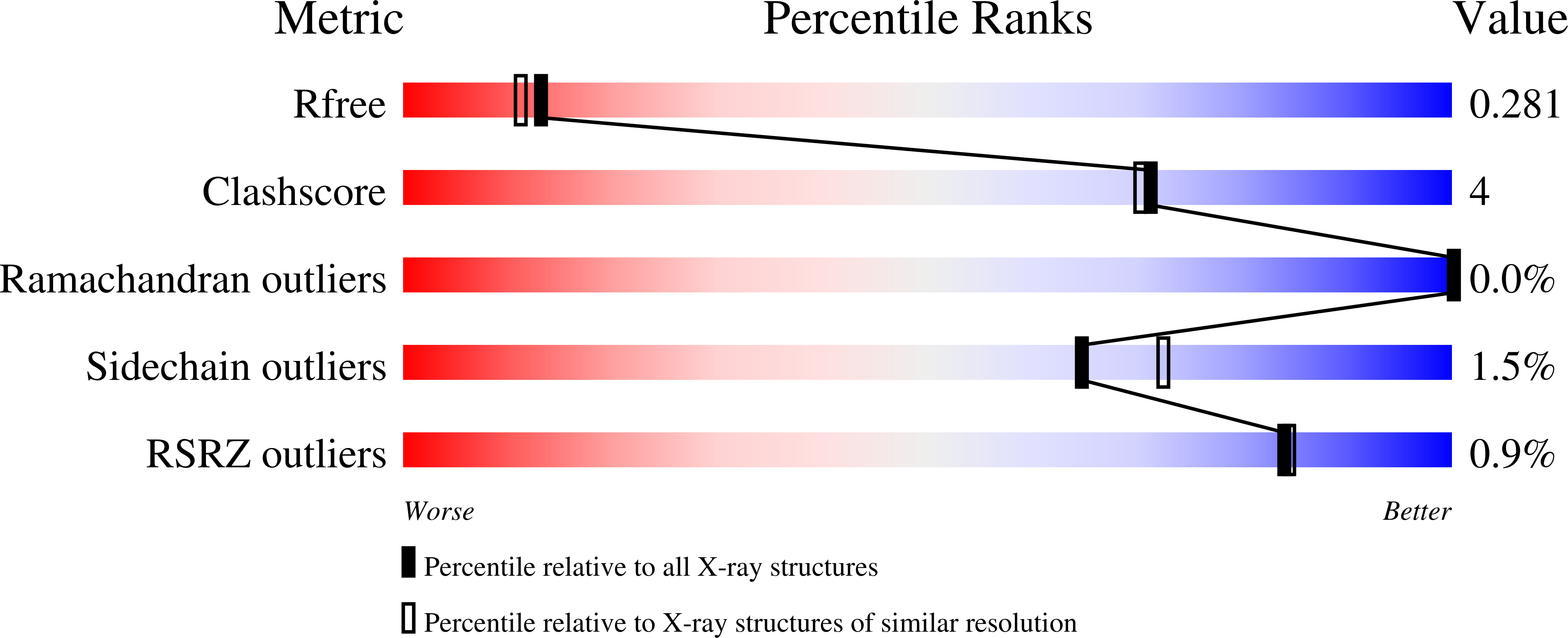
    6X0H, 6X0I, 6X0J, 6X0K

  • PubMed Abstract: 

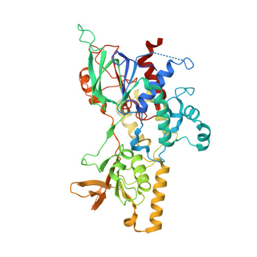
    The siderophore biosynthetic enzyme A (SidA) ornithine hydroxylase from Aspergillus fumigatus is a fungal disease drug target involved in the production of hydroxamate-containing siderophores, which are used by the pathogen to sequester iron. SidA is an N -monooxygenase that catalyzes the NADPH-dependent hydroxylation of l-ornithine through a multistep oxidative mechanism, utilizing a C4a-hydroperoxyflavin intermediate. Here we present four new crystal structures of SidA in various redox and ligation states, including the first structure of oxidized SidA without NADP(H) or l-ornithine bound (resting state). The resting state structure reveals a new out active site conformation characterized by large rotations of the FAD isoalloxazine around the C1-'C2' and N10-C1' bonds, coupled to a 10-Å movement of the Tyr-loop. Additional structures show that either flavin reduction or the binding of NADP(H) is sufficient to drive the FAD to the in conformation. The structures also reveal protein conformational changes associated with the binding of NADP(H) and l-ornithine. Some of these residues were probed using site-directed mutagenesis. Docking was used to explore the active site of the out conformation. These calculations identified two potential ligand-binding sites. Altogether, our results provide new information about conformational dynamics in flavin-dependent monooxygenases. Understanding the different active site conformations that appear during the catalytic cycle may allow fine-tuning of inhibitor discovery efforts.


  • Organizational Affiliation

    Department of Biochemistry, University of Missouri, Columbia, Missouri, USA.


Macromolecules
Find similar proteins by:  (by identity cutoff)  |  3D Structure
Entity ID: 1
MoleculeChains Sequence LengthOrganismDetailsImage
L-ornithine N(5)-monooxygenase
A, B, C, D, E
A, B, C, D, E, F, G, H
494Aspergillus fumigatus Af293Mutation(s): 0 
Gene Names: sidA
EC: 1.14.13.196
UniProt
Find proteins for E9QYP0 (Aspergillus fumigatus (strain ATCC MYA-4609 / CBS 101355 / FGSC A1100 / Af293))
Explore E9QYP0 
Go to UniProtKB:  E9QYP0
Entity Groups  
Sequence Clusters30% Identity50% Identity70% Identity90% Identity95% Identity100% Identity
UniProt GroupE9QYP0
Sequence Annotations
Expand
  • Reference Sequence
Small Molecules
Ligands 2 Unique
IDChains Name / Formula / InChI Key2D Diagram3D Interactions
FDA (Subject of Investigation/LOI)
Query on FDA

Download Ideal Coordinates CCD File 
I [auth A]
K [auth B]
M [auth C]
N [auth D]
P [auth E]
I [auth A],
K [auth B],
M [auth C],
N [auth D],
P [auth E],
R [auth F],
S [auth G],
U [auth H]
DIHYDROFLAVINE-ADENINE DINUCLEOTIDE
C27 H35 N9 O15 P2
YPZRHBJKEMOYQH-UYBVJOGSSA-N
ORN (Subject of Investigation/LOI)
Query on ORN

Download Ideal Coordinates CCD File 
J [auth A]
L [auth B]
O [auth D]
Q [auth E]
T [auth G]
J [auth A],
L [auth B],
O [auth D],
Q [auth E],
T [auth G],
V [auth H]
L-ornithine
C5 H12 N2 O2
AHLPHDHHMVZTML-BYPYZUCNSA-N
Experimental Data & Validation

Experimental Data

  • Method: X-RAY DIFFRACTION
  • Resolution: 2.23 Å
  • R-Value Free: 0.281 
  • R-Value Work: 0.231 
  • R-Value Observed: 0.233 
  • Space Group: P 1 21 1
Unit Cell:
Length ( Å )Angle ( ˚ )
a = 105.902α = 90
b = 155.044β = 91.01
c = 146.846γ = 90
Software Package:
Software NamePurpose
Aimlessdata scaling
PHENIXrefinement
PDB_EXTRACTdata extraction
XDSdata reduction
PHASERphasing

Structure Validation

View Full Validation Report


Ligand Structure Quality Assessment 


Entry History & Funding Information

Deposition Data


Funding OrganizationLocationGrant Number
National Science Foundation (NSF, United States)United StatesCHE-2003658
National Science Foundation (NSF, United States)United StatesCHE-2003986

Revision History  (Full details and data files)

  • Version 1.0: 2020-08-05
    Type: Initial release
  • Version 1.1: 2020-08-12
    Changes: Database references
  • Version 1.2: 2020-09-30
    Changes: Database references
  • Version 1.3: 2023-10-18
    Changes: Data collection, Database references, Refinement description