6WSL

Cryo-EM structure of VASH1-SVBP bound to microtubules


Experimental Data Snapshot

  • Method: ELECTRON MICROSCOPY
  • Resolution: 3.10 Å
  • Aggregation State: FILAMENT 
  • Reconstruction Method: SINGLE PARTICLE 

wwPDB Validation   3D Report Full Report


This is version 1.1 of the entry. See complete history


Literature

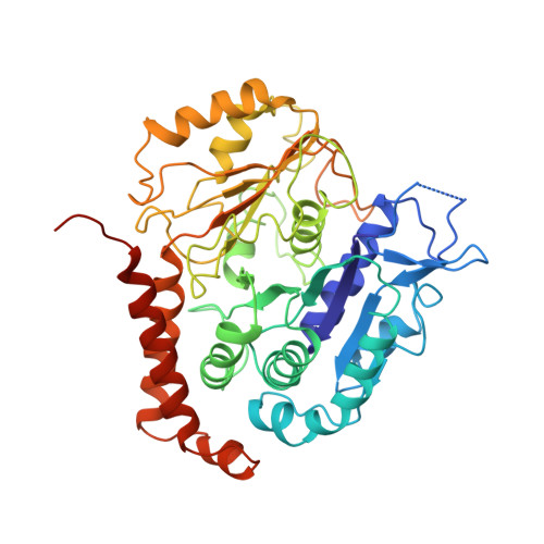
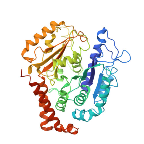
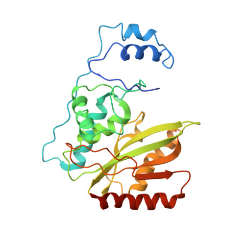
Cryo-EM structure of VASH1-SVBP bound to microtubules.

Li, F.Li, Y.Ye, X.Gao, H.Shi, Z.Luo, X.Rice, L.M.Yu, H.

(2020) Elife 9

  • DOI: https://doi.org/10.7554/eLife.58157
  • Primary Citation of Related Structures:  
    6WSL

  • PubMed Abstract: 

    The dynamic tyrosination-detyrosination cycle of α-tubulin regulates microtubule functions. Perturbation of this cycle impairs mitosis, neural physiology, and cardiomyocyte contraction. The carboxypeptidases vasohibins 1 and 2 (VASH1 and VASH2), in complex with the small vasohibin-binding protein (SVBP), mediate α-tubulin detyrosination. These enzymes detyrosinate microtubules more efficiently than soluble αβ-tubulin heterodimers. The structural basis for this substrate preference is not understood. Using cryo-electron microscopy (cryo-EM), we have determined the structure of human VASH1-SVBP bound to microtubules. The acidic C-terminal tail of α-tubulin binds to a positively charged groove near the active site of VASH1. VASH1 forms multiple additional contacts with the globular domain of α-tubulin, including contacts with a second α-tubulin in an adjacent protofilament. Simultaneous engagement of two protofilaments by VASH1 can only occur within the microtubule lattice, but not with free αβ heterodimers. These lattice-specific interactions enable preferential detyrosination of microtubules by VASH1.


  • Organizational Affiliation

    Department of Pharmacology, University of Texas Southwestern Medical Center, Dallas, United States.


Macromolecules
Find similar proteins by:  (by identity cutoff)  |  3D Structure
Entity ID: 1
MoleculeChains Sequence LengthOrganismDetailsImage
Tubulin alpha-1A chain
A, E
451Homo sapiensMutation(s): 0 
Gene Names: TUBA1ATUBA3
UniProt & NIH Common Fund Data Resources
Find proteins for Q71U36 (Homo sapiens)
Explore Q71U36 
Go to UniProtKB:  Q71U36
PHAROS:  Q71U36
GTEx:  ENSG00000167552 
Entity Groups  
Sequence Clusters30% Identity50% Identity70% Identity90% Identity95% Identity100% Identity
UniProt GroupQ71U36
Sequence Annotations
Expand
  • Reference Sequence
Find similar proteins by:  (by identity cutoff)  |  3D Structure
Entity ID: 2
MoleculeChains Sequence LengthOrganismDetailsImage
Tubulin beta-3 chain
B, F
450Homo sapiensMutation(s): 0 
Gene Names: TUBB3TUBB4
UniProt & NIH Common Fund Data Resources
Find proteins for Q13509 (Homo sapiens)
Explore Q13509 
Go to UniProtKB:  Q13509
PHAROS:  Q13509
GTEx:  ENSG00000258947 
Entity Groups  
Sequence Clusters30% Identity50% Identity70% Identity90% Identity95% Identity100% Identity
UniProt GroupQ13509
Sequence Annotations
Expand
  • Reference Sequence
Find similar proteins by:  (by identity cutoff)  |  3D Structure
Entity ID: 3
MoleculeChains Sequence LengthOrganismDetailsImage
Tubulinyl-Tyr carboxypeptidase 1
C, G
259Homo sapiensMutation(s): 0 
Gene Names: VASH1KIAA1036VASH
EC: 3.4.17.17
UniProt & NIH Common Fund Data Resources
Find proteins for Q7L8A9 (Homo sapiens)
Explore Q7L8A9 
Go to UniProtKB:  Q7L8A9
PHAROS:  Q7L8A9
GTEx:  ENSG00000071246 
Entity Groups  
Sequence Clusters30% Identity50% Identity70% Identity90% Identity95% Identity100% Identity
UniProt GroupQ7L8A9
Sequence Annotations
Expand
  • Reference Sequence
Find similar proteins by:  (by identity cutoff)  |  3D Structure
Entity ID: 4
MoleculeChains Sequence LengthOrganismDetailsImage
Small vasohibin-binding protein
D, H
66Homo sapiensMutation(s): 0 
Gene Names: SVBPCCDC23
UniProt & NIH Common Fund Data Resources
Find proteins for Q8N300 (Homo sapiens)
Explore Q8N300 
Go to UniProtKB:  Q8N300
PHAROS:  Q8N300
GTEx:  ENSG00000177868 
Entity Groups  
Sequence Clusters30% Identity50% Identity70% Identity90% Identity95% Identity100% Identity
UniProt GroupQ8N300
Sequence Annotations
Expand
  • Reference Sequence
Experimental Data & Validation

Experimental Data

  • Method: ELECTRON MICROSCOPY
  • Resolution: 3.10 Å
  • Aggregation State: FILAMENT 
  • Reconstruction Method: SINGLE PARTICLE 

Structure Validation

View Full Validation Report


Entry History & Funding Information

Deposition Data


Funding OrganizationLocationGrant Number
Cancer Prevention and Research Institute of Texas (CPRIT)United StatesRP160667-P2
Welch FoundationUnited StatesI-1441

Revision History  (Full details and data files)

  • Version 1.0: 2020-08-26
    Type: Initial release
  • Version 1.1: 2024-03-06
    Changes: Data collection, Database references