6UKL

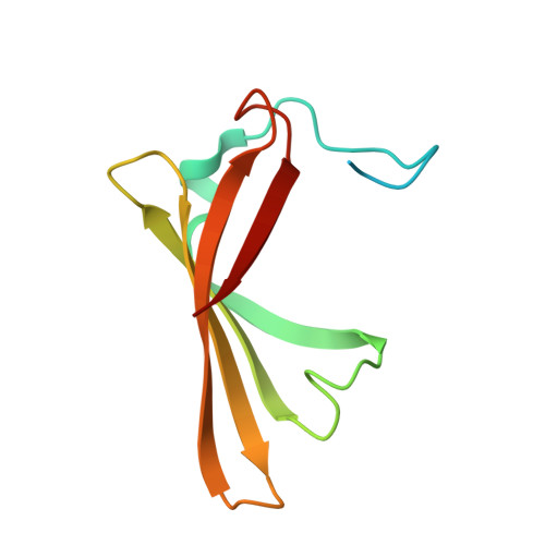
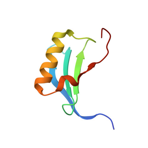
Crystal Structure of a DiB2-split Protein


Experimental Data Snapshot

  • Method: X-RAY DIFFRACTION
  • Resolution: 2.02 Å
  • R-Value Free: 0.248 
  • R-Value Work: 0.205 
  • R-Value Observed: 0.207 

wwPDB Validation   3D Report Full Report


Ligand Structure Quality Assessment 


This is version 1.2 of the entry. See complete history


Literature

DiB-splits: nature-guided design of a novel fluorescent labeling split system.

Bozhanova, N.G.Gavrikov, A.S.Mishin, A.S.Meiler, J.

(2020) Sci Rep 10: 11049-11049

  • DOI: https://doi.org/10.1038/s41598-020-67095-2
  • Primary Citation of Related Structures:  
    6UKK, 6UKL

  • PubMed Abstract: 

    Fluorogen-activating proteins (FAPs) are innovative fluorescent probes combining advantages of genetically-encoded proteins such as green fluorescent protein and externally added fluorogens that allow for highly tunable and on demand fluorescent signaling. Previously, a panel of green- and red-emitting FAPs has been created from bacterial lipocalin Blc (named DiBs). Here we present a rational design as well as functional and structural characterization of the first self-assembling FAP split system, DiB-splits. This new system decreases the size of the FAP label to ~8-12 kDa while preserving DiBs' unique properties: strong increase in fluorescence intensity of the chromophore upon binding, binding affinities to the chromophore in nanomolar to low micromolar range, and high photostability of the protein-ligand complex. These properties allow for use of DiB-splits for wide-field, confocal, and super-resolution fluorescence microscopy. DiB-splits also represent an attractive starting point for further design of a protein-protein interaction detection system as well as novel FAP-based sensors.


  • Organizational Affiliation

    Department of Chemistry, Center for Structural Biology, Vanderbilt University, Nashville, TN, 37235, USA.


Macromolecules
Find similar proteins by:  (by identity cutoff)  |  3D Structure
Entity ID: 1
MoleculeChains Sequence LengthOrganismDetailsImage
Outer membrane lipoprotein BlcA,
B [auth C],
C [auth E]
107Escherichia coliMutation(s): 1 
Gene Names: blcZ5756ECs5130
UniProt
Find proteins for P0A901 (Escherichia coli (strain K12))
Explore P0A901 
Go to UniProtKB:  P0A901
Entity Groups  
Sequence Clusters30% Identity50% Identity70% Identity90% Identity95% Identity100% Identity
UniProt GroupP0A901
Sequence Annotations
Expand
  • Reference Sequence
Find similar proteins by:  (by identity cutoff)  |  3D Structure
Entity ID: 2
MoleculeChains Sequence LengthOrganismDetailsImage
Outer membrane lipoprotein BlcD,
E [auth F],
F [auth B]
69Escherichia coliMutation(s): 0 
Gene Names: blcZ5756ECs5130
UniProt
Find proteins for P0A901 (Escherichia coli (strain K12))
Explore P0A901 
Go to UniProtKB:  P0A901
Entity Groups  
Sequence Clusters30% Identity50% Identity70% Identity90% Identity95% Identity100% Identity
UniProt GroupP0A901
Sequence Annotations
Expand
  • Reference Sequence
Small Molecules
Ligands 3 Unique
IDChains Name / Formula / InChI Key2D Diagram3D Interactions
HEM
Query on HEM

Download Ideal Coordinates CCD File 
R [auth E]PROTOPORPHYRIN IX CONTAINING FE
C34 H32 Fe N4 O4
KABFMIBPWCXCRK-RGGAHWMASA-L
MES
Query on MES

Download Ideal Coordinates CCD File 
O [auth C],
U [auth D]
2-(N-MORPHOLINO)-ETHANESULFONIC ACID
C6 H13 N O4 S
SXGZJKUKBWWHRA-UHFFFAOYSA-N
SO4
Query on SO4

Download Ideal Coordinates CCD File 
G [auth A]
H [auth A]
I [auth A]
J [auth A]
K [auth C]
G [auth A],
H [auth A],
I [auth A],
J [auth A],
K [auth C],
L [auth C],
M [auth C],
N [auth C],
P [auth E],
Q [auth E],
S [auth D],
T [auth D],
V [auth B],
W [auth B],
X [auth B]
SULFATE ION
O4 S
QAOWNCQODCNURD-UHFFFAOYSA-L
Experimental Data & Validation

Experimental Data

  • Method: X-RAY DIFFRACTION
  • Resolution: 2.02 Å
  • R-Value Free: 0.248 
  • R-Value Work: 0.205 
  • R-Value Observed: 0.207 
  • Space Group: P 32 2 1
Unit Cell:
Length ( Å )Angle ( ˚ )
a = 68.241α = 90
b = 68.241β = 90
c = 216.292γ = 120
Software Package:
Software NamePurpose
REFMACrefinement
xia2data scaling
MOLREPphasing
PDB_EXTRACTdata extraction
xia2data reduction

Structure Validation

View Full Validation Report


Ligand Structure Quality Assessment 


Entry History & Funding Information

Deposition Data


Funding OrganizationLocationGrant Number
National Institutes of Health/National Institute of General Medical Sciences (NIH/NIGMS)United StatesR01 GM099842

Revision History  (Full details and data files)

  • Version 1.0: 2020-07-08
    Type: Initial release
  • Version 1.1: 2020-07-22
    Changes: Database references
  • Version 1.2: 2023-10-11
    Changes: Data collection, Database references, Refinement description