6UG0

N2-bound Nitrogenase MoFe-protein from Azotobacter vinelandii


Experimental Data Snapshot

  • Method: X-RAY DIFFRACTION
  • Resolution: 1.83 Å
  • R-Value Free: 0.259 
  • R-Value Work: 0.218 
  • R-Value Observed: 0.218 

wwPDB Validation   3D Report Full Report


Ligand Structure Quality Assessment 


This is version 1.4 of the entry. See complete history


Literature

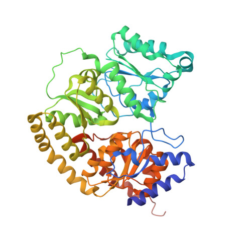
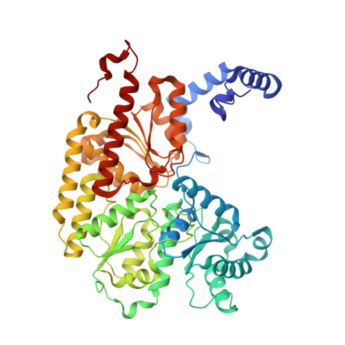
Structural evidence for a dynamic metallocofactor during N2reduction by Mo-nitrogenase.

Kang, W.Lee, C.C.Jasniewski, A.J.Ribbe, M.W.Hu, Y.

(2020) Science 368: 1381-1385

  • DOI: https://doi.org/10.1126/science.aaz6748
  • Primary Citation of Related Structures:  
    6UG0, 6VXT

  • PubMed Abstract: 

    The enzyme nitrogenase uses a suite of complex metallocofactors to reduce dinitrogen (N 2 ) to ammonia. Mechanistic details of this reaction remain sparse. We report a 1.83-angstrom crystal structure of the nitrogenase molybdenum-iron (MoFe) protein captured under physiological N 2 turnover conditions. This structure reveals asymmetric displacements of the cofactor belt sulfurs (S2B or S3A and S5A) with distinct dinitrogen species in the two αβ dimers of the protein. The sulfur-displaced sites are distinct in the ability of protein ligands to donate protons to the bound dinitrogen species, as well as the elongation of either the Mo-O5 (carboxyl) or Mo-O7 (hydroxyl) distance that switches the Mo-homocitrate ligation from bidentate to monodentate. These results highlight the dynamic nature of the cofactor during catalysis and provide evidence for participation of all belt-sulfur sites in this process.


  • Organizational Affiliation

    Department of Molecular Biology and Biochemistry, University of California, Irvine, Irvine, CA 92697-3900, USA.


Macromolecules
Find similar proteins by:  (by identity cutoff)  |  3D Structure
Entity ID: 1
MoleculeChains Sequence LengthOrganismDetailsImage
Nitrogenase molybdenum-iron protein alpha chain
A, C
492Azotobacter vinelandiiMutation(s): 0 
Gene Names: nifD
EC: 1.18.6.1
UniProt
Find proteins for P07328 (Azotobacter vinelandii)
Explore P07328 
Go to UniProtKB:  P07328
Entity Groups  
Sequence Clusters30% Identity50% Identity70% Identity90% Identity95% Identity100% Identity
UniProt GroupP07328
Sequence Annotations
Expand
  • Reference Sequence
Find similar proteins by:  (by identity cutoff)  |  3D Structure
Entity ID: 2
MoleculeChains Sequence LengthOrganismDetailsImage
Nitrogenase molybdenum-iron protein beta chain
B, D
523Azotobacter vinelandiiMutation(s): 0 
Gene Names: nifK
EC: 1.18.6.1
UniProt
Find proteins for P07329 (Azotobacter vinelandii)
Explore P07329 
Go to UniProtKB:  P07329
Entity Groups  
Sequence Clusters30% Identity50% Identity70% Identity90% Identity95% Identity100% Identity
UniProt GroupP07329
Sequence Annotations
Expand
  • Reference Sequence
Small Molecules
Ligands 10 Unique
IDChains Name / Formula / InChI Key2D Diagram3D Interactions
ICE (Subject of Investigation/LOI)
Query on ICE

Download Ideal Coordinates CCD File 
F [auth A]iron-sulfur-molybdenum cluster with interstitial carbon
C Fe7 Mo S8
BJBPMDQXUSSDMI-UHFFFAOYSA-N
ICZ (Subject of Investigation/LOI)
Query on ICZ

Download Ideal Coordinates CCD File 
V [auth C]iron-sulfur-molybdenum cluster with interstitial carbon
C Fe7 Mo S7
BCCRTSRPWONJBV-UHFFFAOYSA-N
1CL (Subject of Investigation/LOI)
Query on 1CL

Download Ideal Coordinates CCD File 
EA [auth C],
L [auth A]
FE(8)-S(7) CLUSTER, OXIDIZED
Fe8 S7
JKVMXLBGZBULKV-UHFFFAOYSA-N
HCA (Subject of Investigation/LOI)
Query on HCA

Download Ideal Coordinates CCD File 
E [auth A],
U [auth C]
3-HYDROXY-3-CARBOXY-ADIPIC ACID
C7 H10 O7
XKJVEVRQMLKSMO-SSDOTTSWSA-N
PGE
Query on PGE

Download Ideal Coordinates CCD File 
R [auth B]TRIETHYLENE GLYCOL
C6 H14 O4
ZIBGPFATKBEMQZ-UHFFFAOYSA-N
MO
Query on MO

Download Ideal Coordinates CCD File 
AA [auth C]
BA [auth C]
I [auth A]
IA [auth D]
J [auth A]
AA [auth C],
BA [auth C],
I [auth A],
IA [auth D],
J [auth A],
K [auth A],
O [auth B],
Z [auth C]
MOLYBDENUM ATOM
Mo
ZOKXTWBITQBERF-UHFFFAOYSA-N
GOL
Query on GOL

Download Ideal Coordinates CCD File 
CA [auth C]
FA [auth D]
GA [auth D]
JA [auth D]
Q [auth B]
CA [auth C],
FA [auth D],
GA [auth D],
JA [auth D],
Q [auth B],
T [auth C]
GLYCEROL
C3 H8 O3
PEDCQBHIVMGVHV-UHFFFAOYSA-N
FE
Query on FE

Download Ideal Coordinates CCD File 
P [auth B],
S [auth B]
FE (III) ION
Fe
VTLYFUHAOXGGBS-UHFFFAOYSA-N
H2S
Query on H2S

Download Ideal Coordinates CCD File 
H [auth A]
HA [auth D]
M [auth B]
N [auth B]
X [auth C]
H [auth A],
HA [auth D],
M [auth B],
N [auth B],
X [auth C],
Y [auth C]
HYDROSULFURIC ACID
H2 S
RWSOTUBLDIXVET-UHFFFAOYSA-N
HDZ (Subject of Investigation/LOI)
Query on HDZ

Download Ideal Coordinates CCD File 
DA [auth C],
G [auth A],
W [auth C]
NITROGEN MOLECULE
N2
IJGRMHOSHXDMSA-UHFFFAOYSA-N
Experimental Data & Validation

Experimental Data

  • Method: X-RAY DIFFRACTION
  • Resolution: 1.83 Å
  • R-Value Free: 0.259 
  • R-Value Work: 0.218 
  • R-Value Observed: 0.218 
  • Space Group: P 21 21 2
Unit Cell:
Length ( Å )Angle ( ˚ )
a = 84.362α = 90
b = 156.967β = 90
c = 202.343γ = 90
Software Package:
Software NamePurpose
Blu-Icedata collection
HKL-3000data reduction
HKL-3000data scaling
PHASERphasing
PHENIXrefinement

Structure Validation

View Full Validation Report


Ligand Structure Quality Assessment 


Entry History & Funding Information

Deposition Data


Funding OrganizationLocationGrant Number
National Institutes of Health/National Institute of General Medical Sciences (NIH/NIGMS)United StatesGM67626
Department of Energy (DOE, United States)United StatesDE-SC0016510

Revision History  (Full details and data files)

  • Version 1.0: 2020-06-24
    Type: Initial release
  • Version 1.1: 2020-07-01
    Changes: Database references
  • Version 1.2: 2020-08-19
    Changes: Derived calculations
  • Version 1.3: 2020-09-30
    Changes: Refinement description
  • Version 1.4: 2023-10-11
    Changes: Advisory, Data collection, Database references, Refinement description