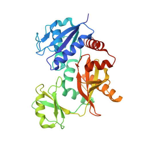
d-Alanine-d-alanine ligase as a model for the activation of ATP-grasp enzymes by monovalent cations.
Pederick, J.L., Thompson, A.P., Bell, S.G., Bruning, J.B.(2020) J Biol Chem 295: 7894-7904
- PubMed: 32335509
- DOI: https://doi.org/10.1074/jbc.RA120.012936
- Primary Citation of Related Structures:
6U1C, 6U1D, 6U1E, 6U1F, 6U1G, 6U1H, 6U1I, 6U1J, 6U1K - PubMed Abstract:
The ATP-grasp superfamily of enzymes shares an atypical nucleotide-binding site known as the ATP-grasp fold. These enzymes are involved in many biological pathways in all domains of life. One ATP-grasp enzyme, d-alanine-d-alanine ligase (Ddl), catalyzes ATP-dependent formation of the d-alanyl-d-alanine dipeptide essential for bacterial cell wall biosynthesis and is therefore an important antibiotic drug target. Ddl is activated by the monovalent cation (MVC) K + , but despite its clinical relevance and decades of research, how this activation occurs has not been elucidated. We demonstrate here that activating MVCs bind adjacent to the active site of Ddl from Thermus thermophilus and used a combined biochemical and structural approach to characterize MVC activation. We found that Tt Ddl is a type II MVC-activated enzyme, retaining activity in the absence of MVCs. However, the efficiency of Tt Ddl increased ∼20-fold in the presence of activating MVCs, and it was maximally activated by K + and Rb + ions. A strict dependence on ionic radius of the MVC was observed, with Li + and Na + providing little to no Tt Ddl activation. To understand the mechanism of MVC activation, we solved crystal structures of Tt Ddl representing distinct catalytic stages in complex with K + , Rb + , or Cs + Comparison of these structures with apo Tt Ddl revealed no evident conformational change on MVC binding. Of note, the identified MVC binding site is structurally conserved within the ATP-grasp superfamily. We propose that MVCs activate Ddl by altering the charge distribution of its active site. These findings provide insight into the catalytic mechanism of ATP-grasp enzymes.
Organizational Affiliation:
Institute for Photonics and Advanced Sensing, School of Biological Sciences, The University of Adelaide, Adelaide, South Australia, Australia.