


Crystal structure of 3'-5' RecJ exonuclease from M. Jannaschii
De March, M., Medagli, B., Krastanova, I., Saha, I., Pisani, F., Onesti, S.To be published.
Experimental Data Snapshot
wwPDB Validation 3D Report Full Report

Entity ID: 1 | |||||
|---|---|---|---|---|---|
| Molecule | Chains | Sequence Length | Organism | Details | Image |
| MjaRecJ | 432 | Methanocaldococcus jannaschii | Mutation(s): 0 Gene Names: MJ0831 | 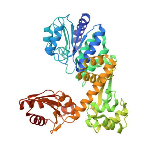 | |
UniProt | |||||
Find proteins for Q58241 (Methanocaldococcus jannaschii (strain ATCC 43067 / DSM 2661 / JAL-1 / JCM 10045 / NBRC 100440)) Explore Q58241 Go to UniProtKB: Q58241 | |||||
Entity Groups | |||||
| Sequence Clusters | 30% Identity50% Identity70% Identity90% Identity95% Identity100% Identity | ||||
| UniProt Group | Q58241 | ||||
Sequence AnnotationsExpand | |||||
| |||||
| Ligands 2 Unique | |||||
|---|---|---|---|---|---|
| ID | Chains | Name / Formula / InChI Key | 2D Diagram | 3D Interactions | |
| GOL Query on GOL | E [auth A], F [auth A], G [auth A] | GLYCEROL C3 H8 O3 PEDCQBHIVMGVHV-UHFFFAOYSA-N |  | ||
| MN (Subject of Investigation/LOI) Query on MN | C [auth A], D [auth A], H [auth B], I [auth B] | MANGANESE (II) ION Mn WAEMQWOKJMHJLA-UHFFFAOYSA-N |  | ||
| Length ( Å ) | Angle ( ˚ ) |
|---|---|
| a = 92.07 | α = 90 |
| b = 92.07 | β = 90 |
| c = 275.76 | γ = 90 |
| Software Name | Purpose |
|---|---|
| REFMAC | refinement |
| XDS | data reduction |
| SCALA | data scaling |
| AutoSol | phasing |
RCSB PDB (citation) is hosted by
RCSB PDB is a member of the

