6TS2

Truncated version of Chaetomium thermophilum UDP-Glucose Glucosyl Transferase (UGGT) lacking domain TRXL2 (417-650).


Experimental Data Snapshot

  • Method: X-RAY DIFFRACTION
  • Resolution: 5.74 Å
  • R-Value Free: 0.249 
  • R-Value Work: 0.174 
  • R-Value Observed: 0.178 

wwPDB Validation   3D Report Full Report


This is version 2.3 of the entry. See complete history


Literature

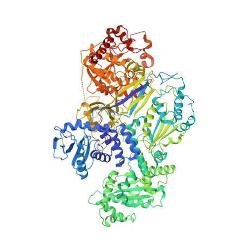
Clamping, bending, and twisting inter-domain motions in the misfold-recognizing portion of UDP-glucose: Glycoprotein glucosyltransferase.

Modenutti, C.P.Blanco Capurro, J.I.Ibba, R.Alonzi, D.S.Song, M.N.Vasiljevic, S.Kumar, A.Chandran, A.V.Tax, G.Marti, L.Hill, J.C.Lia, A.Hensen, M.Waksman, T.Rushton, J.Rubichi, S.Santino, A.Marti, M.A.Zitzmann, N.Roversi, P.

(2021) Structure 29: 357

  • DOI: https://doi.org/10.1016/j.str.2020.11.017
  • Primary Citation of Related Structures:  
    6TRF, 6TRT, 6TS2, 6TS8

  • PubMed Abstract: 

    UDP-glucose:glycoprotein glucosyltransferase (UGGT) flags misfolded glycoproteins for ER retention. We report crystal structures of full-length Chaetomium thermophilum UGGT (CtUGGT), two CtUGGT double-cysteine mutants, and its TRXL2 domain truncation (CtUGGT-ΔTRXL2). CtUGGT molecular dynamics (MD) simulations capture extended conformations and reveal clamping, bending, and twisting inter-domain movements. We name "Parodi limit" the maximum distance on the same glycoprotein between a site of misfolding and an N-linked glycan that can be reglucosylated by monomeric UGGT in vitro, in response to recognition of misfold at that site. Based on the MD simulations, we estimate the Parodi limit as around 70-80 Å. Frequency distributions of distances between glycoprotein residues and their closest N-linked glycosylation sites in glycoprotein crystal structures suggests relevance of the Parodi limit to UGGT activity in vivo. Our data support a "one-size-fits-all adjustable spanner" UGGT substrate recognition model, with an essential role for the UGGT TRXL2 domain.


  • Organizational Affiliation

    Departamento de Química Biológica, Facultad de Ciencias Exactas y Naturales, Universidad de Buenos Aires, Ciudad Universitaria, Pab. II (CE1428EHA), Buenos Aires, Argentina; Instituto de Química Biológica de la Facultad de Ciencias Exactas y Naturales (IQUIBICEN) CONICET. Ciudad Universitaria, Pab. II (CE1428EHA), Buenos Aires, Argentina.


Macromolecules
Find similar proteins by:  (by identity cutoff)  |  3D Structure
Entity ID: 1
MoleculeChains Sequence LengthOrganismDetailsImage
UDP-glucose-glycoprotein glucosyltransferase-like protein,UDP-glucose-glycoprotein glucosyltransferase-like protein
A, B, C, D
1,260Thermochaetoides thermophila DSM 1495Mutation(s): 0 
Gene Names: CTHT_0048990
UniProt
Find proteins for G0SB58 (Chaetomium thermophilum (strain DSM 1495 / CBS 144.50 / IMI 039719))
Explore G0SB58 
Go to UniProtKB:  G0SB58
Entity Groups  
Sequence Clusters30% Identity50% Identity70% Identity90% Identity95% Identity100% Identity
UniProt GroupG0SB58
Sequence Annotations
Expand
  • Reference Sequence
Oligosaccharides

Help

Entity ID: 2
MoleculeChains Length2D Diagram Glycosylation3D Interactions
alpha-D-mannopyranose-(1-3)-[alpha-D-mannopyranose-(1-6)]beta-D-mannopyranose-(1-4)-2-acetamido-2-deoxy-beta-D-glucopyranose-(1-4)-2-acetamido-2-deoxy-beta-D-glucopyranose
E
5N-Glycosylation
Glycosylation Resources
GlyTouCan:  G22768VO
GlyCosmos:  G22768VO
GlyGen:  G22768VO
Entity ID: 3
MoleculeChains Length2D Diagram Glycosylation3D Interactions
beta-D-mannopyranose-(1-4)-2-acetamido-2-deoxy-beta-D-glucopyranose-(1-4)-2-acetamido-2-deoxy-beta-D-glucopyranose
F, H
3N-Glycosylation
Glycosylation Resources
GlyTouCan:  G15407YE
GlyCosmos:  G15407YE
GlyGen:  G15407YE
Entity ID: 4
MoleculeChains Length2D Diagram Glycosylation3D Interactions
alpha-D-mannopyranose-(1-6)-beta-D-mannopyranose-(1-4)-2-acetamido-2-deoxy-beta-D-glucopyranose-(1-4)-2-acetamido-2-deoxy-beta-D-glucopyranose
G
4N-Glycosylation
Glycosylation Resources
GlyTouCan:  G22573RC
GlyCosmos:  G22573RC
GlyGen:  G22573RC
Small Molecules
Ligands 2 Unique
IDChains Name / Formula / InChI Key2D Diagram3D Interactions
NAG
Query on NAG

Download Ideal Coordinates CCD File 
I [auth A]
J [auth A]
K [auth A]
M [auth B]
N [auth B]
I [auth A],
J [auth A],
K [auth A],
M [auth B],
N [auth B],
O [auth B],
Q [auth C],
R [auth C],
S [auth C],
U [auth D],
V [auth D],
W [auth D]
2-acetamido-2-deoxy-beta-D-glucopyranose
C8 H15 N O6
OVRNDRQMDRJTHS-FMDGEEDCSA-N
CA
Query on CA

Download Ideal Coordinates CCD File 
L [auth A],
P [auth B],
T [auth C],
X [auth D]
CALCIUM ION
Ca
BHPQYMZQTOCNFJ-UHFFFAOYSA-N
Experimental Data & Validation

Experimental Data

  • Method: X-RAY DIFFRACTION
  • Resolution: 5.74 Å
  • R-Value Free: 0.249 
  • R-Value Work: 0.174 
  • R-Value Observed: 0.178 
  • Space Group: P 1 21 1
Unit Cell:
Length ( Å )Angle ( ˚ )
a = 151.145α = 90
b = 191.014β = 117.7
c = 158.807γ = 90
Software Package:
Software NamePurpose
BUSTERrefinement
XDSdata reduction
STARANISOdata scaling
MOLREPphasing

Structure Validation

View Full Validation Report


Entry History & Funding Information

Deposition Data


Funding OrganizationLocationGrant Number
Wellcome TrustUnited Kingdom204801/Z/16/Z
Wellcome TrustUnited Kingdom106272/Z/14/Z
Wellcome TrustUnited Kingdom214090/Z/18/Z

Revision History  (Full details and data files)

  • Version 1.0: 2020-01-08
    Type: Initial release
  • Version 2.0: 2020-07-29
    Type: Remediation
    Reason: Carbohydrate remediation
    Changes: Advisory, Atomic model, Data collection, Derived calculations, Structure summary
  • Version 2.1: 2021-01-13
    Changes: Database references, Derived calculations, Structure summary
  • Version 2.2: 2021-04-14
    Changes: Database references
  • Version 2.3: 2024-01-24
    Changes: Data collection, Database references, Refinement description