Functional and structural characterization of PII-like protein CutA does not support involvement in heavy metal tolerance and hints at a small-molecule carrying/signaling role.
Selim, K.A., Tremino, L., Marco-Marin, C., Alva, V., Espinosa, J., Contreras, A., Hartmann, M.D., Forchhammer, K., Rubio, V.(2021) FEBS J 288: 1142-1162
- PubMed: 32599651
- DOI: https://doi.org/10.1111/febs.15464
- Primary Citation of Related Structures:
6GDU, 6GDV, 6GDW, 6GDX, 6T76, 6T7E - PubMed Abstract:
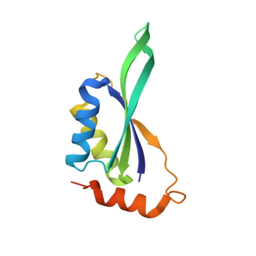
The PII-like protein CutA is annotated as being involved in Cu 2+ tolerance, based on analysis of Escherichia coli mutants. However, the precise cellular function of CutA remains unclear. Our bioinformatic analysis reveals that CutA proteins are universally distributed across all domains of life. Based on sequence-based clustering, we chose representative cyanobacterial CutA proteins for physiological, biochemical, and structural characterization and examined their involvement in heavy metal tolerance, by generating CutA mutants in filamentous Nostoc sp. and in unicellular Synechococcus elongatus. However, we were unable to find any involvement of cyanobacterial CutA in metal tolerance under various conditions. This prompted us to re-examine experimentally the role of CutA in protecting E. coli from Cu 2+ . Since we found no effect on copper tolerance, we conclude that CutA plays a different role that is not involved in metal protection. We resolved high-resolution CutA structures from Nostoc and S. elongatus. Similarly to their counterpart from E. coli and to canonical PII proteins, cyanobacterial CutA proteins are trimeric in solution and in crystal structure; however, no binding affinity for small signaling molecules or for Cu 2+ could be detected. The clefts between the CutA subunits, corresponding to the binding pockets of PII proteins, are formed by conserved aromatic and charged residues, suggesting a conserved binding/signaling function for CutA. In fact, we find binding of organic Bis-Tris/MES molecules in CutA crystal structures, revealing a strong tendency of these pockets to accommodate cargo. This highlights the need to search for the potential physiological ligands and for their signaling functions upon binding to CutA. DATABASES: Structural data are available in Protein Data Bank (PDB) under the accession numbers 6GDU, 6GDV, 6GDW, 6GDX, 6T76, and 6T7E.
Organizational Affiliation:
Interfaculty Institute for Microbiology and Infection Medicine, Organismic Interactions Department, Tübingen University, Germany.