


Structure of Peptide methionine sulfoxide reductase MsrB at 1.63 Angstrom resolution
Napolitano, S., Zyla, D., Glockshuber, R.To be published.
Experimental Data Snapshot
wwPDB Validation 3D Report Full Report

Entity ID: 1 | |||||
|---|---|---|---|---|---|
| Molecule | Chains | Sequence Length | Organism | Details | Image |
| Peptide methionine sulfoxide reductase MsrB | 137 | Escherichia coli K-12 | Mutation(s): 0 Gene Names: msrB, yeaA, b1778, JW1767 EC: 1.8.4.12 | 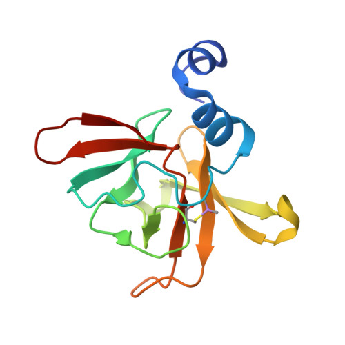 | |
UniProt | |||||
Find proteins for P0A746 (Escherichia coli (strain K12)) Explore P0A746 Go to UniProtKB: P0A746 | |||||
Entity Groups | |||||
| Sequence Clusters | 30% Identity50% Identity70% Identity90% Identity95% Identity100% Identity | ||||
| UniProt Group | P0A746 | ||||
Sequence AnnotationsExpand | |||||
| |||||
| Ligands 1 Unique | |||||
|---|---|---|---|---|---|
| ID | Chains | Name / Formula / InChI Key | 2D Diagram | 3D Interactions | |
| ZN Query on ZN | C [auth A], D [auth B] | ZINC ION Zn PTFCDOFLOPIGGS-UHFFFAOYSA-N |  | ||
| Modified Residues 1 Unique | |||||
|---|---|---|---|---|---|
| ID | Chains | Type | Formula | 2D Diagram | Parent |
| CAF Query on CAF | A, B | L-PEPTIDE LINKING | C5 H12 As N O3 S |  | CYS |
| Length ( Å ) | Angle ( ˚ ) |
|---|---|
| a = 53.39 | α = 90 |
| b = 56.27 | β = 90 |
| c = 85.61 | γ = 90 |
| Software Name | Purpose |
|---|---|
| PHENIX | refinement |
| XDS | data reduction |
| XSCALE | data scaling |
| PHASER | phasing |
| PDB_EXTRACT | data extraction |
RCSB PDB (citation) is hosted by
RCSB PDB is a member of the

