6Q8H

Structure of Fucosylated D-antimicrobial peptide SB10 in complex with the Fucose-binding lectin PA-IIL at 1.707 Angstrom resolution


Experimental Data Snapshot

  • Method: X-RAY DIFFRACTION
  • Resolution: 1.71 Å
  • R-Value Free: 0.189 
  • R-Value Work: 0.168 
  • R-Value Observed: 0.169 

wwPDB Validation   3D Report Full Report


Ligand Structure Quality Assessment 


This is version 2.1 of the entry. See complete history


Literature

X-ray Crystal Structures of Short Antimicrobial Peptides as Pseudomonas aeruginosa Lectin B Complexes.

Baeriswyl, S.Gan, B.H.Siriwardena, T.N.Visini, R.Robadey, M.Javor, S.Stocker, A.Darbre, T.Reymond, J.L.

(2019) ACS Chem Biol 14: 758-766

  • DOI: https://doi.org/10.1021/acschembio.9b00047
  • Primary Citation of Related Structures:  
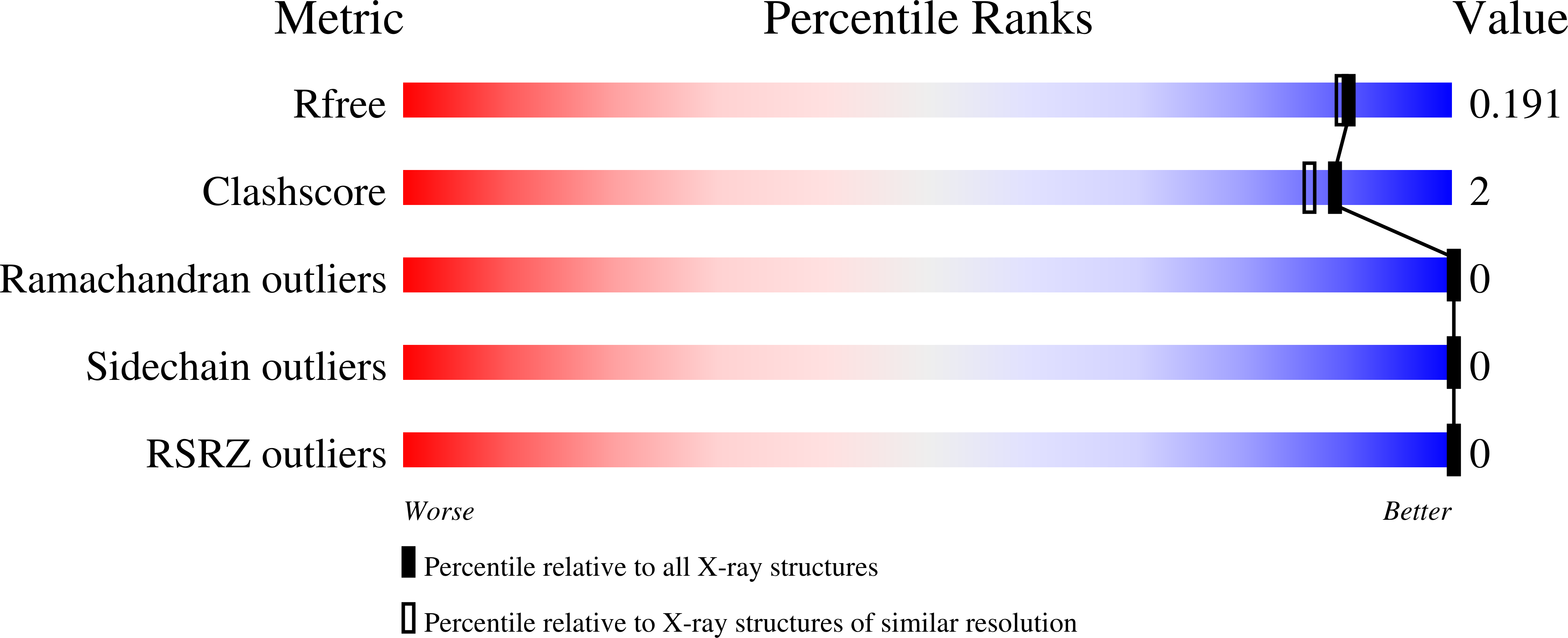
    6Q6W, 6Q6X, 6Q77, 6Q79, 6Q85, 6Q86, 6Q87, 6Q8D, 6Q8G, 6Q8H, 6S5P

  • PubMed Abstract: 

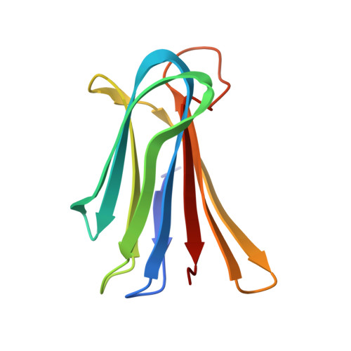
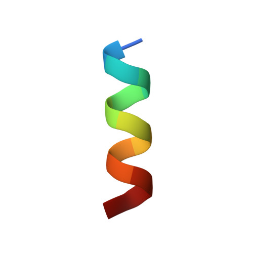
    Herein, we report X-ray crystal structures of 11-13 residue antimicrobial peptides (AMPs) active against Pseudomonas aeruginosa as complexes of fucosylated d-enantiomeric sequences with the P. aeruginosa lectin LecB. These represent the first crystal structures of short AMPs. In 24 individual structures of eight different peptides, we found mostly α-helices assembled as two-helix or four-helix bundles with a hydrophobic core and cationic residues pointing outside. Two of the analogs formed an extended structure engaging in multiple contacts with the lectin. Molecular dynamics (MD) simulations showed that α-helices are stabilized by bundle formation and suggested that the N-terminal acyl group present in the linker to the fucosyl group can extend the helix by one additional H-bond and increase α-helix amphiphilicity. Investigating N-terminal acylation led to AMPs with equivalent and partly stronger antibacterial effects compared to the free peptide.


  • Organizational Affiliation

    Department of Chemistry and Biochemistry , University of Bern , Freiestrasse 3 , 3012 Bern , Switzerland.


Macromolecules
Find similar proteins by:  (by identity cutoff)  |  3D Structure
Entity ID: 1
MoleculeChains Sequence LengthOrganismDetailsImage
Fucose-binding lectin114Pseudomonas aeruginosaMutation(s): 0 
Gene Names: 
UniProt
Find proteins for Q9HYN5 (Pseudomonas aeruginosa (strain ATCC 15692 / DSM 22644 / CIP 104116 / JCM 14847 / LMG 12228 / 1C / PRS 101 / PAO1))
Explore Q9HYN5 
Go to UniProtKB:  Q9HYN5
Entity Groups  
Sequence Clusters30% Identity50% Identity70% Identity90% Identity95% Identity100% Identity
UniProt GroupQ9HYN5
Sequence Annotations
Expand
  • Reference Sequence

Find similar proteins by:  Sequence   |   3D Structure  

Entity ID: 2
MoleculeChains Sequence LengthOrganismDetailsImage
SB10
B, C
13synthetic constructMutation(s): 0 
Sequence Annotations
Expand
  • Reference Sequence
Experimental Data & Validation

Experimental Data

  • Method: X-RAY DIFFRACTION
  • Resolution: 1.71 Å
  • R-Value Free: 0.189 
  • R-Value Work: 0.168 
  • R-Value Observed: 0.169 
  • Space Group: P 62 2 2
Unit Cell:
Length ( Å )Angle ( ˚ )
a = 131.522α = 90
b = 131.522β = 90
c = 49.92γ = 120
Software Package:
Software NamePurpose
PHENIXrefinement
XDSdata reduction
SCALAdata scaling
PHASERphasing
PDB_EXTRACTdata extraction

Structure Validation

View Full Validation Report


Ligand Structure Quality Assessment 


Entry History & Funding Information

Deposition Data


Funding OrganizationLocationGrant Number
Swiss National Science FoundationSwitzerland--

Revision History  (Full details and data files)

  • Version 1.0: 2019-03-20
    Type: Initial release
  • Version 1.1: 2019-05-01
    Changes: Data collection, Database references
  • Version 1.2: 2020-07-29
    Type: Remediation
    Reason: Carbohydrate remediation
    Changes: Data collection, Derived calculations
  • Version 2.0: 2022-12-07
    Changes: Advisory, Atomic model, Data collection, Database references, Derived calculations, Non-polymer description, Polymer sequence, Source and taxonomy, Structure summary
  • Version 2.1: 2024-01-31
    Changes: Data collection, Refinement description