6PBR

Catalytic domain of E.coli dihydrolipoamide succinyltransferase in I4 space group


Experimental Data Snapshot

  • Method: X-RAY DIFFRACTION
  • Resolution: 3.00 Å
  • R-Value Free: 0.272 
  • R-Value Work: 0.230 
  • R-Value Observed: 0.232 

wwPDB Validation   3D Report Full Report


This is version 1.4 of the entry. See complete history


Literature

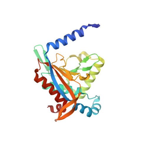
Structure of the dihydrolipoamide succinyltransferase catalytic domain from Escherichia coli in a novel crystal form: a tale of a common protein crystallization contaminant.

Andi, B.Soares, A.S.Shi, W.Fuchs, M.R.McSweeney, S.Liu, Q.

(2019) Acta Crystallogr F Struct Biol Commun 75: 616-624

  • DOI: https://doi.org/10.1107/S2053230X19011488
  • Primary Citation of Related Structures:  
    6PBR

  • PubMed Abstract: 

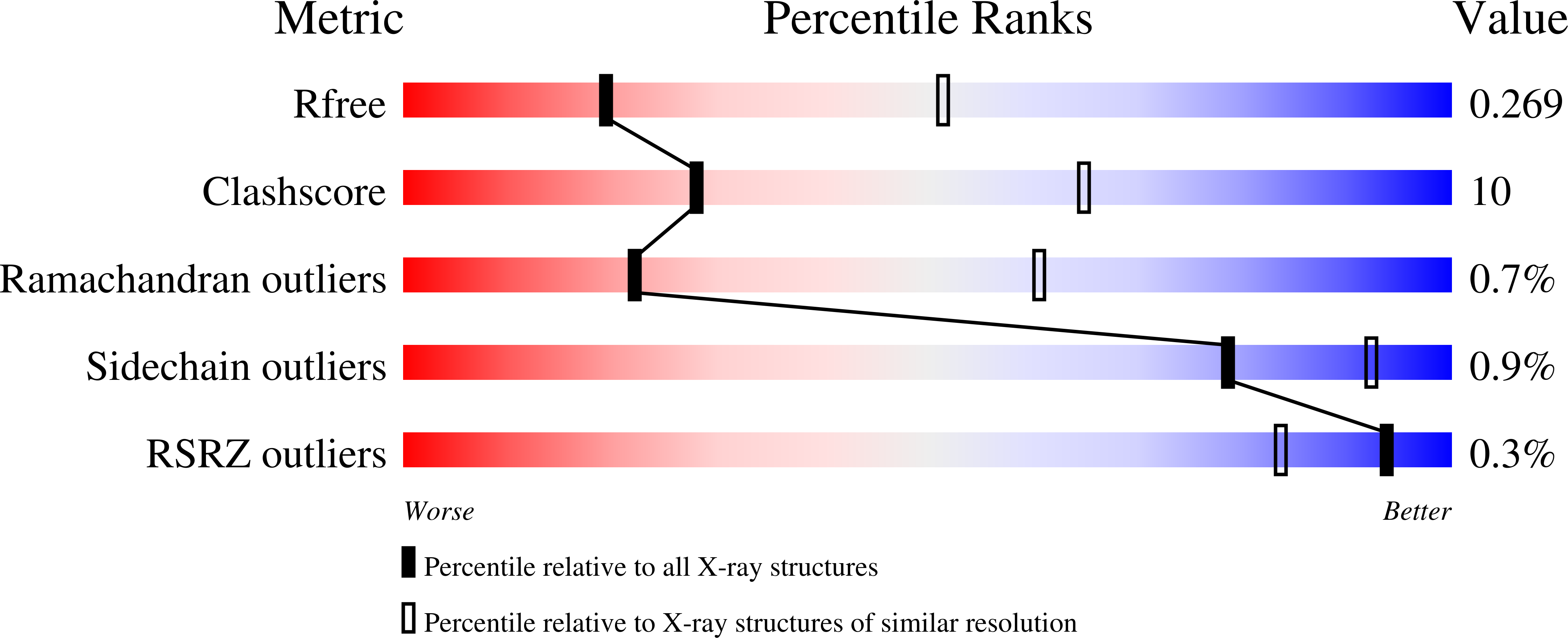
    The crystallization of amidase, the ultimate enzyme in the Trp-dependent auxin-biosynthesis pathway, from Arabidopsis thaliana was attempted using protein samples with at least 95% purity. Cube-shaped crystals that were assumed to be amidase crystals that belonged to space group I4 (unit-cell parameters a = b = 128.6, c = 249.7 Å) were obtained and diffracted to 3.0 Å resolution. Molecular replacement using structures from the PDB containing the amidase signature fold as search models was unsuccessful in yielding a convincing solution. Using the Sequence-Independent Molecular replacement Based on Available Databases (SIMBAD) program, it was discovered that the structure corresponded to dihydrolipoamide succinyltransferase from Escherichia coli (PDB entry 1c4t), which is considered to be a common crystallization contaminant protein. The structure was refined to an R work of 23.0% and an R free of 27.2% at 3.0 Å resolution. The structure was compared with others of the same protein deposited in the PDB. This is the first report of the structure of dihydrolipoamide succinyltransferase isolated without an expression tag and in this novel crystal form.


  • Organizational Affiliation

    National Synchrotron Light Source II, Brookhaven National Laboratory, Upton, NY 11973-5000, USA.


Macromolecules
Find similar proteins by:  (by identity cutoff)  |  3D Structure
Entity ID: 1
MoleculeChains Sequence LengthOrganismDetailsImage
Dihydrolipoyllysine-residue succinyltransferase component of 2-oxoglutarate dehydrogenase complex
A, B, C, D, E
A, B, C, D, E, F
233Escherichia coliMutation(s): 0 
EC: 2.3.1.61
UniProt
Find proteins for P0AFG6 (Escherichia coli (strain K12))
Explore P0AFG6 
Go to UniProtKB:  P0AFG6
Entity Groups  
Sequence Clusters30% Identity50% Identity70% Identity90% Identity95% Identity100% Identity
UniProt GroupP0AFG6
Sequence Annotations
Expand
  • Reference Sequence
Experimental Data & Validation

Experimental Data

  • Method: X-RAY DIFFRACTION
  • Resolution: 3.00 Å
  • R-Value Free: 0.272 
  • R-Value Work: 0.230 
  • R-Value Observed: 0.232 
  • Space Group: I 4
Unit Cell:
Length ( Å )Angle ( ˚ )
a = 128.598α = 90
b = 128.598β = 90
c = 249.733γ = 90
Software Package:
Software NamePurpose
REFMACrefinement
XDSdata reduction
Aimlessdata scaling
SIMBADphasing

Structure Validation

View Full Validation Report


Entry History & Funding Information

Deposition Data


Funding OrganizationLocationGrant Number
National Institutes of Health/National Institute of General Medical Sciences (NIH/NIGMS)United StatesP41GM111244
Department of Energy (DOE, United States)United StatesKP1605010

Revision History  (Full details and data files)

  • Version 1.0: 2019-06-26
    Type: Initial release
  • Version 1.1: 2019-09-11
    Changes: Data collection, Database references
  • Version 1.2: 2019-09-18
    Changes: Data collection, Structure summary
  • Version 1.3: 2019-12-04
    Changes: Author supporting evidence
  • Version 1.4: 2023-10-11
    Changes: Data collection, Database references, Refinement description