6O4P

The crystal structure of the interleukin 11 alpha receptor


Experimental Data Snapshot

  • Method: X-RAY DIFFRACTION
  • Resolution: 3.43 Å
  • R-Value Free: 0.298 
  • R-Value Work: 0.244 
  • R-Value Observed: 0.246 

wwPDB Validation   3D Report Full Report


Ligand Structure Quality Assessment 


This is version 2.0 of the entry. See complete history


Literature

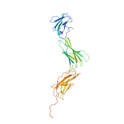
The structure of the extracellular domains of human interleukin 11 alpha receptor reveals mechanisms of cytokine engagement.

Metcalfe, R.D.Aizel, K.Zlatic, C.O.Nguyen, P.M.Morton, C.J.Lio, D.S.Cheng, H.C.Dobson, R.C.J.Parker, M.W.Gooley, P.R.Putoczki, T.L.Griffin, M.D.W.

(2020) J Biol Chem 295: 8285-8301

  • DOI: https://doi.org/10.1074/jbc.RA119.012351
  • Primary Citation of Related Structures:  
    6O4O, 6O4P

  • PubMed Abstract: 

    Interleukin (IL) 11 activates multiple intracellular signaling pathways by forming a complex with its cell surface α-receptor, IL-11Rα, and the β-subunit receptor, gp130. Dysregulated IL-11 signaling has been implicated in several diseases, including some cancers and fibrosis. Mutations in IL-11Rα that reduce signaling are also associated with hereditary cranial malformations. Here we present the first crystal structure of the extracellular domains of human IL-11Rα and a structure of human IL-11 that reveals previously unresolved detail. Disease-associated mutations in IL-11Rα are generally distal to putative ligand-binding sites. Molecular dynamics simulations showed that specific mutations destabilize IL-11Rα and may have indirect effects on the cytokine-binding region. We show that IL-11 and IL-11Rα form a 1:1 complex with nanomolar affinity and present a model of the complex. Our results suggest that the thermodynamic and structural mechanisms of complex formation between IL-11 and IL-11Rα differ substantially from those previously reported for similar cytokines. This work reveals key determinants of the engagement of IL-11 by IL-11Rα that may be exploited in the development of strategies to modulate formation of the IL-11-IL-11Rα complex.


  • Organizational Affiliation

    Department of Biochemistry and Molecular Biology, Bio21 Molecular Science and Biotechnology Institute.


Macromolecules
Find similar proteins by:  (by identity cutoff)  |  3D Structure
Entity ID: 1
MoleculeChains Sequence LengthOrganismDetailsImage
Interleukin-11 receptor subunit alpha
A, B
349Homo sapiensMutation(s): 0 
Gene Names: IL11RA
UniProt & NIH Common Fund Data Resources
Find proteins for Q14626 (Homo sapiens)
Explore Q14626 
Go to UniProtKB:  Q14626
PHAROS:  Q14626
GTEx:  ENSG00000137070 
Entity Groups  
Sequence Clusters30% Identity50% Identity70% Identity90% Identity95% Identity100% Identity
UniProt GroupQ14626
Sequence Annotations
Expand
  • Reference Sequence
Oligosaccharides

Help

Entity ID: 2
MoleculeChains Length2D Diagram Glycosylation3D Interactions
alpha-D-mannopyranose-(1-3)-beta-D-mannopyranose-(1-4)-2-acetamido-2-deoxy-beta-D-glucopyranose-(1-4)-2-acetamido-2-deoxy-beta-D-glucopyranose
C
4N-Glycosylation
Glycosylation Resources
GlyTouCan:  G81315DD
GlyCosmos:  G81315DD
GlyGen:  G81315DD
Entity ID: 3
MoleculeChains Length2D Diagram Glycosylation3D Interactions
2-acetamido-2-deoxy-beta-D-glucopyranose-(1-4)-2-acetamido-2-deoxy-beta-D-glucopyranose
D
2N-Glycosylation
Glycosylation Resources
GlyTouCan:  G42666HT
GlyCosmos:  G42666HT
GlyGen:  G42666HT
Small Molecules
Ligands 2 Unique
IDChains Name / Formula / InChI Key2D Diagram3D Interactions
NAG
Query on NAG

Download Ideal Coordinates CCD File 
E [auth A]2-acetamido-2-deoxy-beta-D-glucopyranose
C8 H15 N O6
OVRNDRQMDRJTHS-FMDGEEDCSA-N
SO4
Query on SO4

Download Ideal Coordinates CCD File 
F [auth A]
G [auth A]
H [auth A]
I [auth A]
J [auth A]
F [auth A],
G [auth A],
H [auth A],
I [auth A],
J [auth A],
K [auth A],
L [auth B],
M [auth B],
N [auth B],
O [auth B],
P [auth B]
SULFATE ION
O4 S
QAOWNCQODCNURD-UHFFFAOYSA-L
Experimental Data & Validation

Experimental Data

  • Method: X-RAY DIFFRACTION
  • Resolution: 3.43 Å
  • R-Value Free: 0.298 
  • R-Value Work: 0.244 
  • R-Value Observed: 0.246 
  • Space Group: P 65 2 2
Unit Cell:
Length ( Å )Angle ( ˚ )
a = 171.456α = 90
b = 171.456β = 90
c = 107.944γ = 120
Software Package:
Software NamePurpose
PHENIXrefinement
XDSdata reduction
XDSdata scaling
PHASERphasing

Structure Validation

View Full Validation Report


Ligand Structure Quality Assessment 


Entry History & Funding Information

Deposition Data


Funding OrganizationLocationGrant Number
National Health and Medical Research Council (NHMRC, Australia)Australia--

Revision History  (Full details and data files)

  • Version 1.0: 2020-05-06
    Type: Initial release
  • Version 1.1: 2020-06-24
    Changes: Database references
  • Version 2.0: 2020-07-29
    Type: Remediation
    Reason: Carbohydrate remediation
    Changes: Atomic model, Data collection, Derived calculations, Structure summary