6M5A

Crystal structure of GH121 beta-L-arabinobiosidase HypBA2 from Bifidobacterium longum


Experimental Data Snapshot

  • Method: X-RAY DIFFRACTION
  • Resolution: 1.85 Å
  • R-Value Free: 0.185 
  • R-Value Work: 0.158 
  • R-Value Observed: 0.159 

wwPDB Validation   3D Report Full Report


This is version 1.2 of the entry. See complete history


Literature

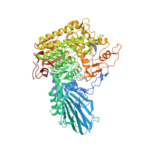
Crystal structure of beta-L-arabinobiosidase belonging to glycoside hydrolase family 121.

Saito, K.Viborg, A.H.Sakamoto, S.Arakawa, T.Yamada, C.Fujita, K.Fushinobu, S.

(2020) PLoS One 15: e0231513-e0231513

  • DOI: https://doi.org/10.1371/journal.pone.0231513
  • Primary Citation of Related Structures:  
    6M5A

  • PubMed Abstract: 

    Enzymes acting on α-L-arabinofuranosides have been extensively studied; however, the structures and functions of β-L-arabinofuranosidases are not fully understood. Three enzymes and an ABC transporter in a gene cluster of Bifidobacterium longum JCM 1217 constitute a degradation and import system of β-L-arabinooligosaccharides on plant hydroxyproline-rich glycoproteins. An extracellular β-L-arabinobiosidase (HypBA2) belonging to the glycoside hydrolase (GH) family 121 plays a key role in the degradation pathway by releasing β-1,2-linked arabinofuranose disaccharide (β-Ara2) for the specific sugar importer. Here, we present the crystal structure of the catalytic region of HypBA2 as the first three-dimensional structure of GH121 at 1.85 Å resolution. The HypBA2 structure consists of a central catalytic (α/α)6 barrel domain and two flanking (N- and C-terminal) β-sandwich domains. A pocket in the catalytic domain appears to be suitable for accommodating the β-Ara2 disaccharide. Three acidic residues Glu383, Asp515, and Glu713, located in this pocket, are completely conserved among all members of GH121; site-directed mutagenesis analysis showed that they are essential for catalytic activity. The active site of HypBA2 was compared with those of structural homologs in other GH families: GH63 α-glycosidase, GH94 chitobiose phosphorylase, GH142 β-L-arabinofuranosidase, GH78 α-L-rhamnosidase, and GH37 α,α-trehalase. Based on these analyses, we concluded that the three conserved residues are essential for catalysis and substrate binding. β-L-Arabinobiosidase genes in GH121 are mainly found in the genomes of bifidobacteria and Xanthomonas species, suggesting that the cleavage and specific import system for the β-Ara2 disaccharide on plant hydroxyproline-rich glycoproteins are shared in animal gut symbionts and plant pathogens.


  • Organizational Affiliation

    Department of Biotechnology, The University of Tokyo, Tokyo, Japan.


Macromolecules
Find similar proteins by:  (by identity cutoff)  |  3D Structure
Entity ID: 1
MoleculeChains Sequence LengthOrganismDetailsImage
Beta-L-arabinobiosidase871Bifidobacterium longumMutation(s): 0 
Gene Names: DWV93_04865
UniProt
Find proteins for E8MGH9 (Bifidobacterium longum subsp. longum (strain ATCC 15707 / DSM 20219 / JCM 1217 / NCTC 11818 / E194b))
Explore E8MGH9 
Go to UniProtKB:  E8MGH9
Entity Groups  
Sequence Clusters30% Identity50% Identity70% Identity90% Identity95% Identity100% Identity
UniProt GroupE8MGH9
Sequence Annotations
Expand
  • Reference Sequence
Small Molecules
Ligands 5 Unique
IDChains Name / Formula / InChI Key2D Diagram3D Interactions
PG4
Query on PG4

Download Ideal Coordinates CCD File 
F [auth A]TETRAETHYLENE GLYCOL
C8 H18 O5
UWHCKJMYHZGTIT-UHFFFAOYSA-N
PGE
Query on PGE

Download Ideal Coordinates CCD File 
C [auth A],
H [auth A],
P [auth A],
X [auth A]
TRIETHYLENE GLYCOL
C6 H14 O4
ZIBGPFATKBEMQZ-UHFFFAOYSA-N
PEG
Query on PEG

Download Ideal Coordinates CCD File 
B [auth A]
E [auth A]
G [auth A]
I [auth A]
L [auth A]
B [auth A],
E [auth A],
G [auth A],
I [auth A],
L [auth A],
N [auth A],
S [auth A],
T [auth A]
DI(HYDROXYETHYL)ETHER
C4 H10 O3
MTHSVFCYNBDYFN-UHFFFAOYSA-N
EDO
Query on EDO

Download Ideal Coordinates CCD File 
D [auth A]
J [auth A]
K [auth A]
M [auth A]
O [auth A]
D [auth A],
J [auth A],
K [auth A],
M [auth A],
O [auth A],
Q [auth A],
R [auth A],
U [auth A],
V [auth A],
W [auth A]
1,2-ETHANEDIOL
C2 H6 O2
LYCAIKOWRPUZTN-UHFFFAOYSA-N
CA
Query on CA

Download Ideal Coordinates CCD File 
AA [auth A],
Y [auth A],
Z [auth A]
CALCIUM ION
Ca
BHPQYMZQTOCNFJ-UHFFFAOYSA-N
Experimental Data & Validation

Experimental Data

  • Method: X-RAY DIFFRACTION
  • Resolution: 1.85 Å
  • R-Value Free: 0.185 
  • R-Value Work: 0.158 
  • R-Value Observed: 0.159 
  • Space Group: P 21 21 21
Unit Cell:
Length ( Å )Angle ( ˚ )
a = 81.526α = 90
b = 88.616β = 90
c = 127.918γ = 90
Software Package:
Software NamePurpose
REFMACrefinement
PDB_EXTRACTdata extraction
XDSdata reduction
Aimlessdata scaling
Aimlessphasing

Structure Validation

View Full Validation Report


Entry History & Funding Information

Deposition Data


Funding OrganizationLocationGrant Number
Japan Society for the Promotion of Science (JSPS)Japan24380053
Japan Society for the Promotion of Science (JSPS)Japan22780094
Japan Society for the Promotion of Science (JSPS)Japan26660083
Japan Society for the Promotion of Science (JSPS)Japan15H02443

Revision History  (Full details and data files)

  • Version 1.0: 2020-06-03
    Type: Initial release
  • Version 1.1: 2020-07-01
    Changes: Database references
  • Version 1.2: 2024-03-27
    Changes: Data collection, Database references