6JU1

p-Hydroxybenzoate hydroxylase Y385F mutant complexed with 3,4-dihydroxybenzoate


Experimental Data Snapshot

  • Method: X-RAY DIFFRACTION
  • Resolution: 1.60 Å
  • R-Value Free: 0.186 
  • R-Value Work: 0.158 
  • R-Value Observed: 0.159 

wwPDB Validation   3D Report Full Report


Ligand Structure Quality Assessment 


This is version 1.2 of the entry. See complete history


Literature

Understanding the Molecular Mechanism Underlying the High Catalytic Activity ofp-Hydroxybenzoate Hydroxylase Mutants for Producing Gallic Acid.

Moriwaki, Y.Yato, M.Terada, T.Saito, S.Nukui, N.Iwasaki, T.Nishi, T.Kawaguchi, Y.Okamoto, K.Arakawa, T.Yamada, C.Fushinobu, S.Shimizu, K.

(2019) Biochemistry 58: 4543-4558

  • DOI: https://doi.org/10.1021/acs.biochem.9b00443
  • Primary Citation of Related Structures:  
    6JU1

  • PubMed Abstract: 

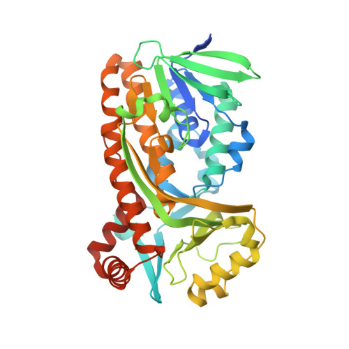
    p -Hydroxybenzoate hydroxylase (PHBH) is a flavoprotein monooxygenase that catalyzes the hydroxylation of p -hydroxybenzoate ( p -OHB) to 3,4-dihydroxybenzoate (3,4-DOHB). PHBH can bind to other benzoate derivatives in addition to p -OHB; however, hydroxylation does not occur on 3,4-DOHB. Replacement of Tyr385 with Phe forms a mutant, which enables the production of 3,4,5-trihydroxybenzonate (gallic acid) from 3,4-DOHB, although the catalytic activity of the mutant is quite low. In this study, we report how the L199V/Y385F double mutant exhibits activity for producing gallic acid 4.3-fold higher than that of the Y385F single mutant. This improvement in catalytic activity is primarily due to the suppression of a shunt reaction that wastes reduced nicotinamide adenine dinucleotide phosphate by producing H 2 O 2 . To further elucidate the molecular mechanism underlying this higher catalytic activity, we performed molecular dynamics simulations and quantum mechanics/molecular mechanics calculations, in addition to determining the crystal structure of the Y385F·3,4-DOHB complex. The simulations showed that the Y385F mutation facilitates the deprotonation of the 4-hydroxy group of 3,4-DOHB, which is necessary for initiating hydroxylation. Moreover, the L199V mutation in addition to the Y385F mutation allows the OH moiety in the peroxide group of C-(4a)-flavin hydroperoxide to come into the proximity of the C5 atom of 3,4-DOHB. Overall, this study provides a consistent explanation for the change in the catalytic activity of PHBH caused by mutations, which will enable us to better design an enzyme with different activities.


  • Organizational Affiliation

    The Collaborative Research Institute for Innovative Microbiology , The University of Tokyo , 1-1-1 Yayoi , Bunkyo-ku, Tokyo 113-8657 , Japan.


Macromolecules
Find similar proteins by:  (by identity cutoff)  |  3D Structure
Entity ID: 1
MoleculeChains Sequence LengthOrganismDetailsImage
4-hydroxybenzoate 3-monooxygenase414Pseudomonas aeruginosaMutation(s): 1 
Gene Names: pobADT376_03235
EC: 1.14.13.2
UniProt
Find proteins for P20586 (Pseudomonas aeruginosa (strain ATCC 15692 / DSM 22644 / CIP 104116 / JCM 14847 / LMG 12228 / 1C / PRS 101 / PAO1))
Explore P20586 
Go to UniProtKB:  P20586
Entity Groups  
Sequence Clusters30% Identity50% Identity70% Identity90% Identity95% Identity100% Identity
UniProt GroupP20586
Sequence Annotations
Expand
  • Reference Sequence
Small Molecules
Ligands 6 Unique
IDChains Name / Formula / InChI Key2D Diagram3D Interactions
FAD (Subject of Investigation/LOI)
Query on FAD

Download Ideal Coordinates CCD File 
B [auth A]FLAVIN-ADENINE DINUCLEOTIDE
C27 H33 N9 O15 P2
VWWQXMAJTJZDQX-UYBVJOGSSA-N
P33
Query on P33

Download Ideal Coordinates CCD File 
I [auth A]3,6,9,12,15,18-HEXAOXAICOSANE-1,20-DIOL
C14 H30 O8
XPJRQAIZZQMSCM-UHFFFAOYSA-N
PG4
Query on PG4

Download Ideal Coordinates CCD File 
H [auth A]TETRAETHYLENE GLYCOL
C8 H18 O5
UWHCKJMYHZGTIT-UHFFFAOYSA-N
DHB (Subject of Investigation/LOI)
Query on DHB

Download Ideal Coordinates CCD File 
C [auth A]3,4-DIHYDROXYBENZOIC ACID
C7 H6 O4
YQUVCSBJEUQKSH-UHFFFAOYSA-N
GOL
Query on GOL

Download Ideal Coordinates CCD File 
D [auth A],
E [auth A],
F [auth A],
G [auth A]
GLYCEROL
C3 H8 O3
PEDCQBHIVMGVHV-UHFFFAOYSA-N
BR
Query on BR

Download Ideal Coordinates CCD File 
J [auth A],
K [auth A]
BROMIDE ION
Br
CPELXLSAUQHCOX-UHFFFAOYSA-M
Experimental Data & Validation

Experimental Data

  • Method: X-RAY DIFFRACTION
  • Resolution: 1.60 Å
  • R-Value Free: 0.186 
  • R-Value Work: 0.158 
  • R-Value Observed: 0.159 
  • Space Group: C 2 2 21
Unit Cell:
Length ( Å )Angle ( ˚ )
a = 70.866α = 90
b = 145.901β = 90
c = 86.421γ = 90
Software Package:
Software NamePurpose
REFMACrefinement
HKL-2000data reduction
HKL-2000data scaling
MOLREPphasing

Structure Validation

View Full Validation Report


Ligand Structure Quality Assessment 


Entry History 

Deposition Data

Revision History  (Full details and data files)

  • Version 1.0: 2019-11-06
    Type: Initial release
  • Version 1.1: 2019-11-20
    Changes: Database references
  • Version 1.2: 2023-11-22
    Changes: Data collection, Database references, Refinement description, Structure summary