Structural Basis of Mitochondrial Scaffolds by Prohibitin Complexes: Insight into a Role of the Coiled-Coil Region.
Yoshinaka, T., Kosako, H., Yoshizumi, T., Furukawa, R., Hirano, Y., Kuge, O., Tamada, T., Koshiba, T.(2019) iScience 19: 1065-1078
- PubMed: 31522117
- DOI: https://doi.org/10.1016/j.isci.2019.08.056
- Primary Citation of Related Structures:
6IQE - PubMed Abstract:
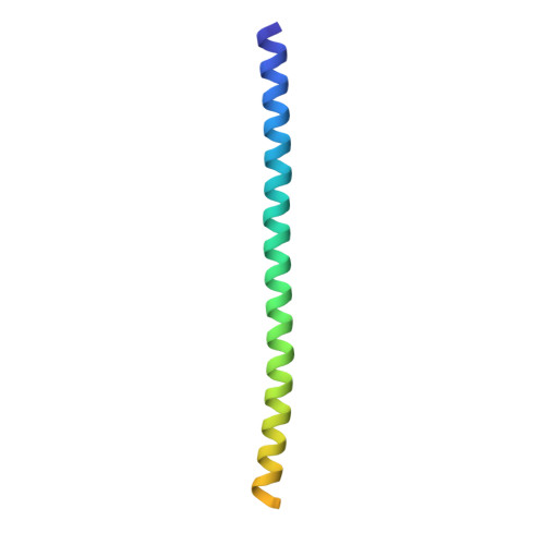
The coiled-coil motif mediates subunit oligomerization and scaffolding and underlies several fundamental biologic processes. Prohibitins (PHBs), mitochondrial inner membrane proteins involved in mitochondrial homeostasis and signal transduction, are predicted to have a coiled-coil motif, but their structural features are poorly understood. Here we solved the crystal structure of the heptad repeat (HR) region of PHB2 at 1.7-Å resolution, showing that it assembles into a dimeric, antiparallel coiled-coil with a unique negatively charged area essential for the PHB interactome in mitochondria. Disruption of the HR coiled-coil abolishes well-ordered PHB complexes and the mitochondrial tubular networks accompanying PHB-dependent signaling. Using a proximity-dependent biotin identification (BioID) technique in live cells, we mapped a number of mitochondrial intermembrane space proteins whose association with PHB2 relies on the HR coiled-coil region. Elucidation of the PHB complex structure in mitochondria provides insight into essential PHB interactomes required for mitochondrial dynamics as well as signal transduction.
Organizational Affiliation:
Department of Biology, Faculty of Science, Kyushu University, Fukuoka 819-0395, Japan.