6IDJ

Crystal structure of human DHODH in complex with ferulenol


Experimental Data Snapshot

  • Method: X-RAY DIFFRACTION
  • Resolution: 1.90 Å
  • R-Value Free: 0.178 
  • R-Value Work: 0.163 
  • R-Value Observed: 0.163 

wwPDB Validation   3D Report Full Report


Ligand Structure Quality Assessment 


This is version 1.2 of the entry. See complete history


Literature

Structural and Biochemical Features of Eimeria tenella Dihydroorotate Dehydrogenase, a Potential Drug Target.

Sato, D.Hartuti, E.D.Inaoka, D.K.Sakura, T.Amalia, E.Nagahama, M.Yoshioka, Y.Tsuji, N.Nozaki, T.Kita, K.Harada, S.Matsubayashi, M.Shiba, T.

(2020) Genes (Basel) 11

  • DOI: https://doi.org/10.3390/genes11121468
  • Primary Citation of Related Structures:  
    6AJ5, 6AJE, 6IDJ

  • PubMed Abstract: 

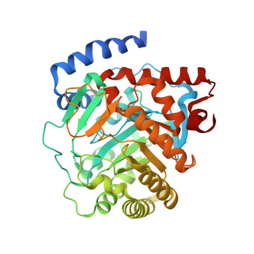
    Dihydroorotate dehydrogenase (DHODH) is a mitochondrial monotopic membrane protein that plays an essential role in the pyrimidine de novo biosynthesis and electron transport chain pathways. In Eimeria tenella , an intracellular apicomplexan parasite that causes the most severe form of chicken coccidiosis, the activity of pyrimidine salvage pathway at the intracellular stage is negligible and it relies on the pyrimidine de novo biosynthesis pathway. Therefore, the enzymes of the de novo pathway are considered potential drug target candidates for the design of compounds with activity against this parasite. Although, DHODHs from E. tenella (EtDHODH), Plasmodium falciparum (PfDHODH), and human (HsDHODH) show distinct sensitivities to classical DHODH inhibitors, in this paper, we identify ferulenol as a potent inhibitor of both EtDHODH and HsDHODH. Additionally, we report the crystal structures of EtDHODH and HsDHODH in the absence and presence of ferulenol. Comparison of these enzymes showed that despite similar overall structures, the EtDHODH has a long insertion in the N-terminal helix region that assumes a disordered configuration. In addition, the crystal structures revealed that the ferulenol binding pocket of EtDHODH is larger than that of HsDHODH. These differences can be explored to accelerate structure-based design of inhibitors specifically targeting EtDHODH.


  • Organizational Affiliation

    Department of Applied Biology, Graduate School of Science Technology, Kyoto Institute of Technology, Matsugasaki, Sakyo-ku, Kyoto 606-8585, Japan.


Macromolecules
Find similar proteins by:  (by identity cutoff)  |  3D Structure
Entity ID: 1
MoleculeChains Sequence LengthOrganismDetailsImage
Dihydroorotate dehydrogenase (quinone), mitochondrial390Homo sapiensMutation(s): 0 
Gene Names: DHODH
EC: 1.3.5.2
UniProt & NIH Common Fund Data Resources
Find proteins for Q02127 (Homo sapiens)
Explore Q02127 
Go to UniProtKB:  Q02127
PHAROS:  Q02127
GTEx:  ENSG00000102967 
Entity Groups  
Sequence Clusters30% Identity50% Identity70% Identity90% Identity95% Identity100% Identity
UniProt GroupQ02127
Sequence Annotations
Expand
  • Reference Sequence
Small Molecules
Ligands 5 Unique
IDChains Name / Formula / InChI Key2D Diagram3D Interactions
FMN
Query on FMN

Download Ideal Coordinates CCD File 
E [auth A]FLAVIN MONONUCLEOTIDE
C17 H21 N4 O9 P
FVTCRASFADXXNN-SCRDCRAPSA-N
9AU
Query on 9AU

Download Ideal Coordinates CCD File 
G [auth A]4-oxidanyl-3-[(2~{E},6~{E})-3,7,11-trimethyldodeca-2,6,10-trienyl]chromen-2-one
C24 H30 O3
NJJDBBUWWOAOLD-CFBAGHHKSA-N
ORO
Query on ORO

Download Ideal Coordinates CCD File 
F [auth A]OROTIC ACID
C5 H4 N2 O4
PXQPEWDEAKTCGB-UHFFFAOYSA-N
SO4
Query on SO4

Download Ideal Coordinates CCD File 
B [auth A],
H [auth A],
I [auth A]
SULFATE ION
O4 S
QAOWNCQODCNURD-UHFFFAOYSA-L
ACT
Query on ACT

Download Ideal Coordinates CCD File 
C [auth A],
D [auth A],
J [auth A]
ACETATE ION
C2 H3 O2
QTBSBXVTEAMEQO-UHFFFAOYSA-M
Experimental Data & Validation

Experimental Data

  • Method: X-RAY DIFFRACTION
  • Resolution: 1.90 Å
  • R-Value Free: 0.178 
  • R-Value Work: 0.163 
  • R-Value Observed: 0.163 
  • Space Group: P 32 2 1
Unit Cell:
Length ( Å )Angle ( ˚ )
a = 90.155α = 90
b = 90.155β = 90
c = 123.334γ = 120
Software Package:
Software NamePurpose
REFMACrefinement
HKL-2000data reduction
HKL-2000data scaling
MOLREPphasing

Structure Validation

View Full Validation Report


Ligand Structure Quality Assessment 


Entry History 

Deposition Data

Revision History  (Full details and data files)

  • Version 1.0: 2019-09-11
    Type: Initial release
  • Version 1.1: 2020-12-23
    Changes: Database references
  • Version 1.2: 2023-11-22
    Changes: Data collection, Database references, Refinement description