6I8W

Crystal structure of a membrane phospholipase A, a novel bacterial virulence factor


Experimental Data Snapshot

  • Method: X-RAY DIFFRACTION
  • Resolution: 2.00 Å
  • R-Value Free: 0.186 
  • R-Value Work: 0.163 
  • R-Value Observed: 0.164 

wwPDB Validation   3D Report Full Report


Ligand Structure Quality Assessment 


This is version 1.5 of the entry. See complete history


Literature

Structural, mechanistic, and physiological insights into phospholipase A-mediated membrane phospholipid degradation in Pseudomonas aeruginosa.

Bleffert, F.Granzin, J.Caliskan, M.Schott-Verdugo, S.N.Siebers, M.Thiele, B.Rahme, L.Felgner, S.Dormann, P.Gohlke, H.Batra-Safferling, R.Jaeger, K.E.Kovacic, F.

(2022) Elife 11

  • DOI: https://doi.org/10.7554/eLife.72824
  • Primary Citation of Related Structures:  
    6I8W

  • PubMed Abstract: 

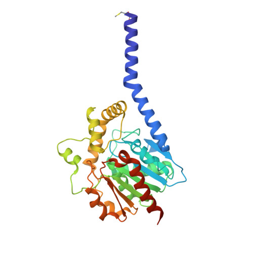
    Cells steadily adapt their membrane glycerophospholipid (GPL) composition to changing environmental and developmental conditions. While the regulation of membrane homeostasis via GPL synthesis in bacteria has been studied in detail, the mechanisms underlying the controlled degradation of endogenous GPLs remain unknown. Thus far, the function of intracellular phospholipases A (PLAs) in GPL remodeling (Lands cycle) in bacteria is not clearly established. Here, we identified the first cytoplasmic membrane-bound phospholipase A 1 (PlaF) from Pseudomonas aeruginosa , which might be involved in the Lands cycle. PlaF is an important virulence factor, as the P. aeruginosa Δ plaF mutant showed strongly attenuated virulence in Galleria mellonella and macrophages. We present a 2.0-Å-resolution crystal structure of PlaF, the first structure that reveals homodimerization of a single-pass transmembrane (TM) full-length protein. PlaF dimerization, mediated solely through the intermolecular interactions of TM and juxtamembrane regions, inhibits its activity. The dimerization site and the catalytic sites are linked by an intricate ligand-mediated interaction network, which might explain the product (fatty acid) feedback inhibition observed with the purified PlaF protein. We used molecular dynamics simulations and configurational free energy computations to suggest a model of PlaF activation through a coupled monomerization and tilting of the monomer in the membrane, which constrains the active site cavity into contact with the GPL substrates. Thus, these data show the importance of the PlaF-mediated GPL remodeling pathway for virulence and could pave the way for the development of novel therapeutics targeting PlaF.


  • Organizational Affiliation

    Institute of Molecular Enzyme Technology, Heinrich Heine University Düsseldorf, Forschungszentrum Jülich GmbH, Jülich, Germany.


Macromolecules
Find similar proteins by:  (by identity cutoff)  |  3D Structure
Entity ID: 1
MoleculeChains Sequence LengthOrganismDetailsImage
Alpha/beta fold hydrolase
A, B
322Pseudomonas aeruginosaMutation(s): 0 
Gene Names: lip3AOY09_04297C8257_11100DN070_25360NCTC13719_02038PAMH19_2121
EC: 3.1.1.3
Membrane Entity: Yes 
UniProt
Find proteins for Q9KJG6 (Pseudomonas aeruginosa)
Explore Q9KJG6 
Go to UniProtKB:  Q9KJG6
Entity Groups  
Sequence Clusters30% Identity50% Identity70% Identity90% Identity95% Identity100% Identity
UniProt GroupQ9KJG6
Sequence Annotations
Expand
  • Reference Sequence
Small Molecules
Ligands 5 Unique
IDChains Name / Formula / InChI Key2D Diagram3D Interactions
BOG
Query on BOG

Download Ideal Coordinates CCD File 
D [auth A],
J [auth B]
octyl beta-D-glucopyranoside
C14 H28 O6
HEGSGKPQLMEBJL-RKQHYHRCSA-N
MYR
Query on MYR

Download Ideal Coordinates CCD File 
C [auth A]MYRISTIC ACID
C14 H28 O2
TUNFSRHWOTWDNC-UHFFFAOYSA-N
11A
Query on 11A

Download Ideal Coordinates CCD File 
I [auth B]UNDECANOIC ACID
C11 H22 O2
ZDPHROOEEOARMN-UHFFFAOYSA-N
IPA
Query on IPA

Download Ideal Coordinates CCD File 
E [auth A],
K [auth B],
L [auth B]
ISOPROPYL ALCOHOL
C3 H8 O
KFZMGEQAYNKOFK-UHFFFAOYSA-N
CO2
Query on CO2

Download Ideal Coordinates CCD File 
F [auth A],
G [auth A],
H [auth A],
M [auth B],
N [auth B]
CARBON DIOXIDE
C O2
CURLTUGMZLYLDI-UHFFFAOYSA-N
Modified Residues  1 Unique
IDChains TypeFormula2D DiagramParent
FME
Query on FME
A, B
L-PEPTIDE LINKINGC6 H11 N O3 SMET
Experimental Data & Validation

Experimental Data

  • Method: X-RAY DIFFRACTION
  • Resolution: 2.00 Å
  • R-Value Free: 0.186 
  • R-Value Work: 0.163 
  • R-Value Observed: 0.164 
  • Space Group: I 41 2 2
Unit Cell:
Length ( Å )Angle ( ˚ )
a = 133.871α = 90
b = 133.871β = 90
c = 212.358γ = 90
Software Package:
Software NamePurpose
PHENIXrefinement
XDSdata reduction
Aimlessdata scaling
MrBUMPphasing

Structure Validation

View Full Validation Report


Ligand Structure Quality Assessment 


Entry History 

Deposition Data

Revision History  (Full details and data files)

  • Version 1.0: 2019-11-27
    Type: Initial release
  • Version 1.1: 2020-06-10
    Changes: Database references
  • Version 1.2: 2020-07-29
    Type: Remediation
    Reason: Carbohydrate remediation
    Changes: Data collection, Derived calculations, Structure summary
  • Version 1.3: 2022-05-18
    Changes: Database references, Structure summary
  • Version 1.4: 2023-04-26
    Changes: Database references, Structure summary
  • Version 1.5: 2024-02-07
    Changes: Data collection, Refinement description