6HMJ

Structure of an RNA-binding Light-Oxygen-Voltage Receptor


Experimental Data Snapshot

  • Method: X-RAY DIFFRACTION
  • Resolution: 2.51 Å
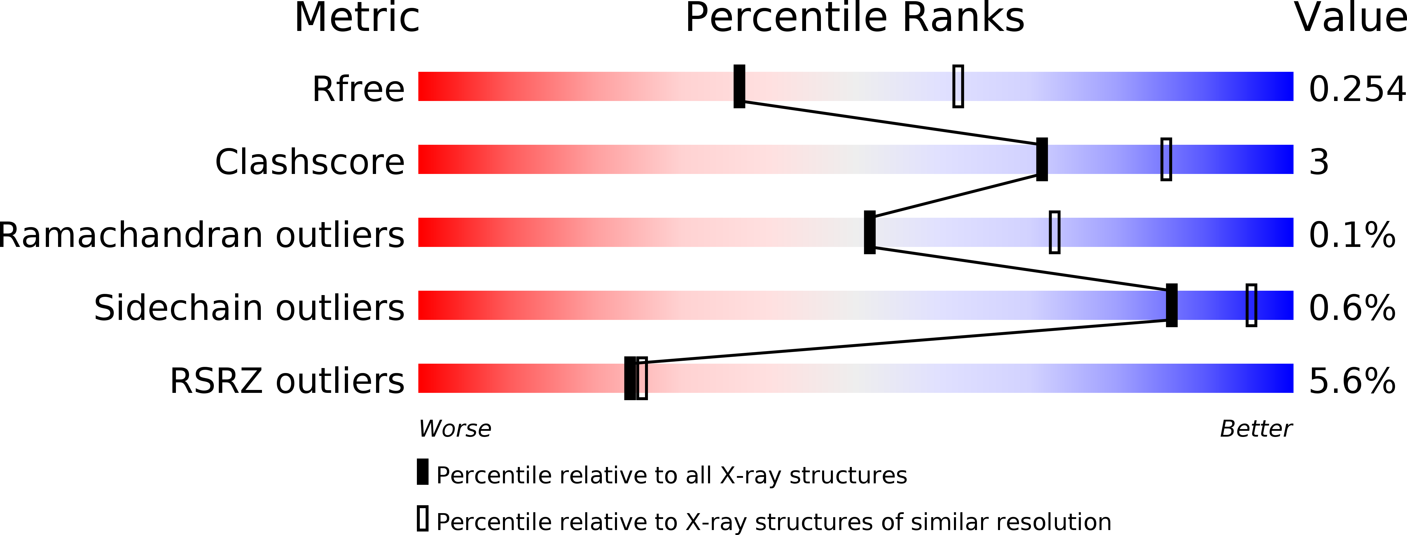
  • R-Value Free: 0.254 
  • R-Value Work: 0.243 
  • R-Value Observed: 0.244 

wwPDB Validation   3D Report Full Report


Ligand Structure Quality Assessment 


This is version 1.1 of the entry. See complete history


Literature

A blue light receptor that mediates RNA binding and translational regulation.

Weber, A.M.Kaiser, J.Ziegler, T.Pilsl, S.Renzl, C.Sixt, L.Pietruschka, G.Moniot, S.Kakoti, A.Juraschitz, M.Schrottke, S.Lledo Bryant, L.Steegborn, C.Bittl, R.Mayer, G.Moglich, A.

(2019) Nat Chem Biol 15: 1085-1092

  • DOI: https://doi.org/10.1038/s41589-019-0346-y
  • Primary Citation of Related Structures:  
    6HMJ

  • PubMed Abstract: 

    Sensory photoreceptor proteins underpin light-dependent adaptations in nature and enable the optogenetic control of organismal behavior and physiology. We identified the bacterial light-oxygen-voltage (LOV) photoreceptor PAL that sequence-specifically binds short RNA stem loops with around 20 nM affinity in blue light and weaker than 1 µM in darkness. A crystal structure rationalizes the unusual receptor architecture of PAL with C-terminal LOV photosensor and N-terminal effector units. The light-activated PAL-RNA interaction can be harnessed to regulate gene expression at the RNA level as a function of light in both bacteria and mammalian cells. The present results elucidate a new signal-transduction paradigm in LOV receptors and conjoin RNA biology with optogenetic regulation, thereby paving the way toward hitherto inaccessible optoribogenetic modalities.


  • Organizational Affiliation

    LIMES Institut, Universität Bonn, Bonn, Germany.


Macromolecules
Find similar proteins by:  (by identity cutoff)  |  3D Structure
Entity ID: 1
MoleculeChains Sequence LengthOrganismDetailsImage
Putative PAS/PAC sensor protein
A, B, C, D
373Nakamurella multipartita DSM 44233Mutation(s): 0 
Gene Names: Namu_4361
UniProt
Find proteins for C8XJT7 (Nakamurella multipartita (strain ATCC 700099 / DSM 44233 / CIP 104796 / JCM 9543 / NBRC 105858 / Y-104))
Explore C8XJT7 
Go to UniProtKB:  C8XJT7
Entity Groups  
Sequence Clusters30% Identity50% Identity70% Identity90% Identity95% Identity100% Identity
UniProt GroupC8XJT7
Sequence Annotations
Expand
  • Reference Sequence
Small Molecules
Ligands 4 Unique
IDChains Name / Formula / InChI Key2D Diagram3D Interactions
FMN
Query on FMN

Download Ideal Coordinates CCD File 
E [auth A],
G [auth B],
I [auth C],
K [auth D]
FLAVIN MONONUCLEOTIDE
C17 H21 N4 O9 P
FVTCRASFADXXNN-SCRDCRAPSA-N
GOL
Query on GOL

Download Ideal Coordinates CCD File 
F [auth A],
L [auth D],
N [auth D]
GLYCEROL
C3 H8 O3
PEDCQBHIVMGVHV-UHFFFAOYSA-N
IMD
Query on IMD

Download Ideal Coordinates CCD File 
H [auth B]IMIDAZOLE
C3 H5 N2
RAXXELZNTBOGNW-UHFFFAOYSA-O
EDO
Query on EDO

Download Ideal Coordinates CCD File 
J [auth C],
M [auth D]
1,2-ETHANEDIOL
C2 H6 O2
LYCAIKOWRPUZTN-UHFFFAOYSA-N
Experimental Data & Validation

Experimental Data

  • Method: X-RAY DIFFRACTION
  • Resolution: 2.51 Å
  • R-Value Free: 0.254 
  • R-Value Work: 0.243 
  • R-Value Observed: 0.244 
  • Space Group: P 21 21 21
Unit Cell:
Length ( Å )Angle ( ˚ )
a = 67.013α = 90
b = 150.345β = 90
c = 219.783γ = 90
Software Package:
Software NamePurpose
PHENIXrefinement
DIALSdata reduction
Aimlessdata scaling
SHELXCDphasing

Structure Validation

View Full Validation Report


Ligand Structure Quality Assessment 


Entry History & Funding Information

Deposition Data


Funding OrganizationLocationGrant Number
German Research FoundationGermanyMO2192/6-1

Revision History  (Full details and data files)

  • Version 1.0: 2019-08-28
    Type: Initial release
  • Version 1.1: 2020-09-09
    Changes: Database references