6HCP

Crystal structure of BauA, the Ferric preacinetobactin receptor from Acinetobacter baumannii


Experimental Data Snapshot

  • Method: X-RAY DIFFRACTION
  • Resolution: 1.83 Å
  • R-Value Free: 0.177 
  • R-Value Work: 0.156 
  • R-Value Observed: 0.157 

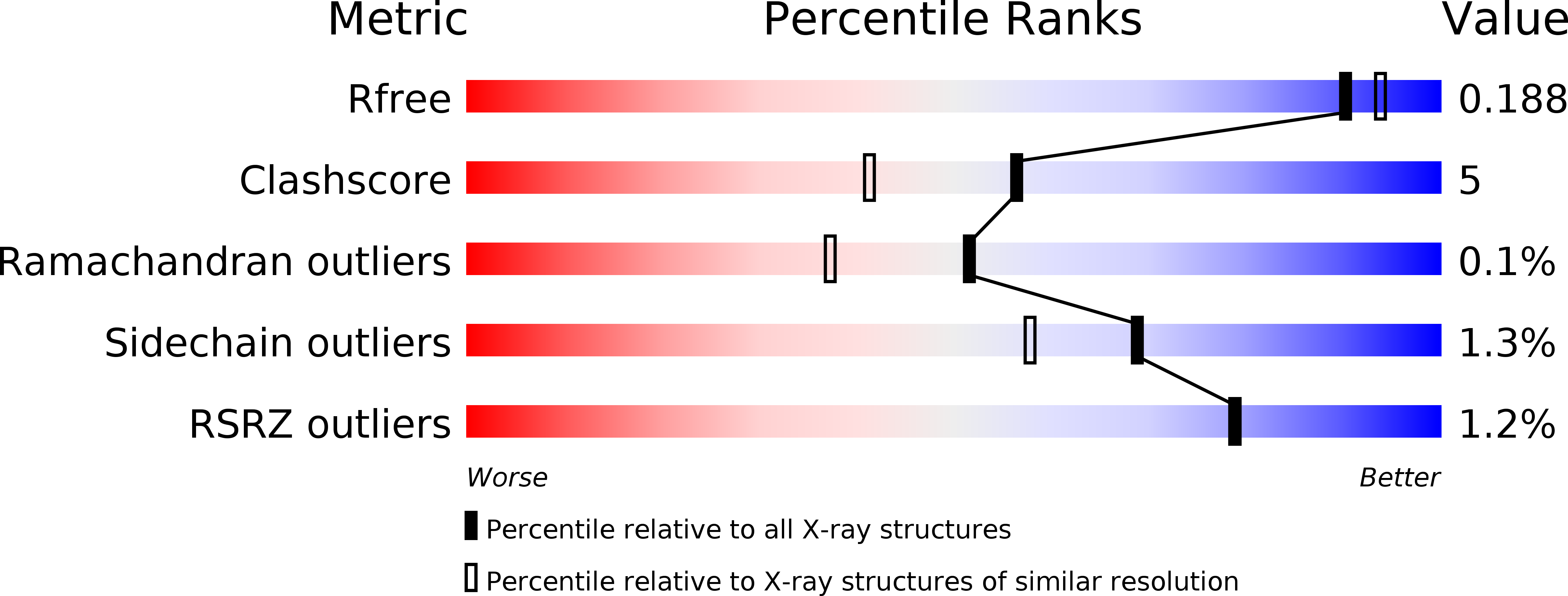
wwPDB Validation   3D Report Full Report


Ligand Structure Quality Assessment 


This is version 1.1 of the entry. See complete history


Literature

Preacinetobactin not acinetobactin is essential for iron uptake by the BauA transporter of the pathogenAcinetobacter baumannii.

Moynie, L.Serra, I.Scorciapino, M.A.Oueis, E.Page, M.G.Ceccarelli, M.Naismith, J.H.

(2018) Elife 7

  • DOI: https://doi.org/10.7554/eLife.42270
  • Primary Citation of Related Structures:  
    6H7F, 6H7V, 6HCP

  • PubMed Abstract: 

    New strategies are urgently required to develop antibiotics. The siderophore uptake system has attracted considerable attention, but rational design of siderophore antibiotic conjugates requires knowledge of recognition by the cognate outer-membrane transporter. Acinetobacter baumannii is a serious pathogen, which utilizes (pre)acinetobactin to scavenge iron from the host. We report the structure of the (pre)acinetobactin transporter BauA bound to the siderophore, identifying the structural determinants of recognition. Detailed biophysical analysis confirms that BauA recognises preacinetobactin. We show that acinetobactin is not recognised by the protein, thus preacinetobactin is essential for iron uptake. The structure shows and NMR confirms that under physiological conditions, a molecule of acinetobactin will bind to two free coordination sites on the iron preacinetobactin complex. The ability to recognise a heterotrimeric iron-preacinetobactin-acinetobactin complex may rationalize contradictory reports in the literature. These results open new avenues for the design of novel antibiotic conjugates (trojan horse) antibiotics.


  • Organizational Affiliation

    Division of Structural Biology, Wellcome Trust Centre of Human Genomics, Oxford, England.


Macromolecules
Find similar proteins by:  (by identity cutoff)  |  3D Structure
Entity ID: 1
MoleculeChains Sequence LengthOrganismDetailsImage
BauA
A, B, C
706Acinetobacter baumanniiMutation(s): 0 
Gene Names: bauA
Membrane Entity: Yes 
UniProt
Find proteins for Q76HJ9 (Acinetobacter baumannii)
Explore Q76HJ9 
Go to UniProtKB:  Q76HJ9
Entity Groups  
Sequence Clusters30% Identity50% Identity70% Identity90% Identity95% Identity100% Identity
UniProt GroupQ76HJ9
Sequence Annotations
Expand
  • Reference Sequence
Small Molecules
Ligands 3 Unique
IDChains Name / Formula / InChI Key2D Diagram3D Interactions
C8E
Query on C8E

Download Ideal Coordinates CCD File 
AD [auth C]
BD [auth C]
CA [auth A]
CD [auth C]
DA [auth A]
AD [auth C],
BD [auth C],
CA [auth A],
CD [auth C],
DA [auth A],
DD [auth C],
EA [auth A],
ED [auth C],
FA [auth A],
FD [auth C],
GA [auth A],
HA [auth A],
IA [auth A],
JA [auth A],
KA [auth A],
LA [auth A],
LB [auth B],
MA [auth A],
MB [auth B],
NB [auth B],
OB [auth B],
PB [auth B],
QB [auth B],
RB [auth B],
SB [auth B],
SC [auth C],
TB [auth B],
TC [auth C],
UB [auth B],
UC [auth C],
VC [auth C],
WC [auth C],
XC [auth C],
YC [auth C],
ZC [auth C]
(HYDROXYETHYLOXY)TRI(ETHYLOXY)OCTANE
C16 H34 O5
FEOZZFHAVXYAMB-UHFFFAOYSA-N
PO4
Query on PO4

Download Ideal Coordinates CCD File 
VB [auth B]PHOSPHATE ION
O4 P
NBIIXXVUZAFLBC-UHFFFAOYSA-K
EDO
Query on EDO

Download Ideal Coordinates CCD File 
AA [auth A]
AB [auth B]
AC [auth C]
BA [auth A]
BB [auth B]
AA [auth A],
AB [auth B],
AC [auth C],
BA [auth A],
BB [auth B],
BC [auth C],
CB [auth B],
CC [auth C],
D [auth A],
DB [auth B],
DC [auth C],
E [auth A],
EB [auth B],
EC [auth C],
F [auth A],
FB [auth B],
FC [auth C],
G [auth A],
GB [auth B],
GC [auth C],
H [auth A],
HB [auth B],
HC [auth C],
I [auth A],
IB [auth B],
IC [auth C],
J [auth A],
JB [auth B],
JC [auth C],
K [auth A],
KB [auth B],
KC [auth C],
L [auth A],
LC [auth C],
M [auth A],
MC [auth C],
N [auth A],
NA [auth B],
NC [auth C],
O [auth A],
OA [auth B],
OC [auth C],
P [auth A],
PA [auth B],
PC [auth C],
Q [auth A],
QA [auth B],
QC [auth C],
R [auth A],
RA [auth B],
RC [auth C],
S [auth A],
SA [auth B],
T [auth A],
TA [auth B],
U [auth A],
UA [auth B],
V [auth A],
VA [auth B],
W [auth A],
WA [auth B],
WB [auth C],
X [auth A],
XA [auth B],
XB [auth C],
Y [auth A],
YA [auth B],
YB [auth C],
Z [auth A],
ZA [auth B],
ZB [auth C]
1,2-ETHANEDIOL
C2 H6 O2
LYCAIKOWRPUZTN-UHFFFAOYSA-N
Experimental Data & Validation

Experimental Data

  • Method: X-RAY DIFFRACTION
  • Resolution: 1.83 Å
  • R-Value Free: 0.177 
  • R-Value Work: 0.156 
  • R-Value Observed: 0.157 
  • Space Group: C 1 2 1
Unit Cell:
Length ( Å )Angle ( ˚ )
a = 180.2α = 90
b = 219.52β = 99.21
c = 101.43γ = 90
Software Package:
Software NamePurpose
REFMACrefinement
XDSdata reduction
XSCALEdata scaling
PHASERphasing

Structure Validation

View Full Validation Report


Ligand Structure Quality Assessment 


Entry History & Funding Information

Deposition Data


Funding OrganizationLocationGrant Number
United Kingdom--

Revision History  (Full details and data files)

  • Version 1.0: 2018-10-10
    Type: Initial release
  • Version 1.1: 2019-01-16
    Changes: Data collection, Database references, Structure summary